| Description |
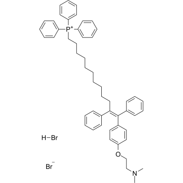
MitoTam bromide, hydrobromide is a tamoxifen derivative[1], an electron transport chain (ETC) inhibitor, spreduces mitochondrial membrane potential in senescent cells and affects mitochondrial morphology[2].MitoTam bromide, hydrobromide is an effective anticancer agent, suppresses respiratory complexes (CI-respiration) and disrupts respiratory supercomplexes (SCs) formation in breast cancer cells[1][2]. MitoTam bromide, hydrobromide causes apoptosis[2].
|
| In Vitro |
MitoTam (0.5 μM-56 μM; 24 hours) kills breast cancer cell Lines and nonmalignant cells with an IC50 range from 0.65 μM to 55.9 μM[1]. MitoTam (2.5 μM; 2-24 hours) results in stronger activation of the apoptotic pathway in MCF7 Her2high cells compared with mock MCF7 cells[1].MitoTam (0.05 μM-1 μM; 3 days) causes a concentration-dependent induction of apoptosis in breast cancer cells, while there was no effect for non-malignant breast epithelial cells[2] Cell Viability Assay[1] Cell Line: Breast Cancer Cell Lines: BT474, MCF7, MCF7 Her2high, MCF7 Her2low, MDA-MB-231, MDA-MB-436, MDA-MB-453, SK-BR-3, T47D; NeuTL cells; Nonmalignant Cells: A014578, H9c2 cells Concentration: 0.5 μM-56 μM Incubation Time: 24 hours Result: Killed breast cancer cells MCF7, MCF7 Her2high, MCF7 Her2low with IC50 values of 1.25 μM, 0.65 μM and 1.45 μM respectively. Western Blot Analysis[1] Cell Line: MCF7 mock cells, MCF7 Her2high cells Concentration: 2.5 μM Incubation Time: 2 hours, 4 hours, 8 hours, 16 hours, 24 hours Result: Revealed accelerated cleavage of procaspase-9, Parp1/2 and proapoptotic Bax, decreased the antiapoptotic Bcl-2 protein in Her2high cells. Apoptosis Analysis[2] Cell Line: MCF-7 cells, 4T1 cells and MCF-10a cells Concentration: 0.05 μM-1 μM Incubation Time: 3 days Result: Resulted in apoptosis in MCF7 and 4T1 cells.
|
