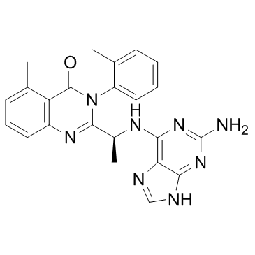
CAL-130
Modify Date: 2024-01-09 11:40:52

CAL-130 structure
|
Common Name | CAL-130 | ||
|---|---|---|---|---|
| CAS Number | 1431697-74-3 | Molecular Weight | 426.47 | |
| Density | N/A | Boiling Point | N/A | |
| Molecular Formula | C23H22N8O | Melting Point | N/A | |
| MSDS | N/A | Flash Point | N/A | |
Use of CAL-130CAL-130 is a PI3Kδ and PI3Kγ inhibitor with IC50s of 1.3 and 6.1 nM, respectively. |
| Name | CAL 130 |
|---|---|
| Synonym | More Synonyms |
| Description | CAL-130 is a PI3Kδ and PI3Kγ inhibitor with IC50s of 1.3 and 6.1 nM, respectively. |
|---|---|
| Related Catalog | |
| Target |
p110δ:1.3 nM (IC50) p110γ:6.1 nM (IC50) p110β:56 nM (IC50) p110α:115 nM (IC50) |
| In Vitro | CAL-130 preferentially inhibits the function of both p110γ and p110δ catalytic domains. IC50 values of CAL-130 are 1.3 and 6.1 nM for p110δ and p110γ, respectively, as compared to 115 and 56 nM for p110α and p110β. CAL-130 does not inhibit additional intracellular signaling pathways (i.e., p38 MAPK or insulin receptor tyrosine kinase) that are critical for general cell function and survival[1]. |
| In Vivo | The clinical significance of interfering with the combined activities of PI3Kγ and PI3Kδ is determined by administering CAL-130 to Lck/Ptenfl/fl mice with established T cell acute lymphoblastic leukemia (T-ALL). Candidate animals for survival studies are ill appearing, have a white blood cell (WBC) count above 45,000 μL-1, evidence of blasts on peripheral smear, and a majority of circulation cells (>75%) staining double positive for Thy1.2 and Ki-67. Mice receive an oral dose (10 mg/kg) of CAL-130 every 8 hr for a period of 7 days and are then followed until moribund. Despite the limited duration of therapy, CAL-130 is highly effective in extending the median survival for treated animals to 45 days as compared 7.5 days for the control group[1]. |
| Kinase Assay | IC50 values for CAL-130 inhibition of PI3K isoforms are determined in ex vivo PI3 kinase assays using recombinant PI3K. A ten-point kinase inhibitory profile is determined with ATP at a concentration consistent with the KM for each enzyme[1]. |
| Cell Assay | Cell proliferation of CCRF-CEM cells or shRNA-transfected CCRF-CEM cells, in presence or absence of CAL-130 (1, 2.5 and 5 μM), is followed by cell counting of samples in triplicate using a hemocytometer and trypan blue. For apoptosis determinations of untransfected or shRNA-transfected CCRF-CEMs, cells are stained with APC-conjugated Annexin-V in Annexin Binding Buffer and analyzed by flow cytometry. For primary T-ALL samples, cell viability is assessed using the BD Cell Viability kit coupled with the use of fluorescentcounting beads. For this, cells are plated with MS5-DL1 stroma cells, and after 72 hr following CAL-130 treatment, cells are harvested and stained with an APC-conjugated antihuman CD45 followed by a staining with the aforementioned kit[1]. |
| Animal Admin | Mice[1] For subcutaneous xenograft experiments, luminescent CCRF-CEM (CEMluc) cells are generated by lentiviral infection with FUW-luc and selection with Neomycin. Luciferase expression is verified with the Dual-Luciferase Reporter Assay kit. 2.5×10⁶ CEM-luc cells embedded in Matrigel are injected in the flank of NOD.Cg-Prkdcscid Il2rgtm1Wjl/Sz mice. After 1 week, mice are treated by oral gavage with vehicle (0.5% methyl cellulose, 0.1% Tween 80), or CAL-130 (10 mg/kg) every 8 hr daily for 4 days, and then tumors are imaged as follows: mice anesthetized by isoflurane inhalation are injected intraperitoneally with D-luciferin (50 mg/kg). Photonic emission is imaged with the in vivo imaging system. Tumor bioluminescence is quantified by integrating the photonic flux (photons per second) through a region encircling each tumor using the Living Image software package. Administration of D-luciferin and detection of tumor bioluminescence in Lck/Ptenfl/fl/Gt(ROSA)26Sortm1(Luc)Kael/J mice are performed in a similar manner. |
| References |
| Molecular Formula | C23H22N8O |
|---|---|
| Molecular Weight | 426.47 |
| Storage condition | 2-8℃ |
| CAL-130 |