| Description |
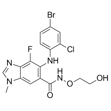
Selumetinib is a highly potent MEK inhibitor, with an IC50 of 14 nM against MEK1.
|
| Related Catalog |
|
| Target |
MEK:12 nM (IC50)
MEK1:14 nM (IC50)
|
| In Vitro |
Selumetinib causes a time- and dose-dependent reduction in DNA synthesis and cell viability in primary, induces growth arrest and apoptosis associated with the inactivation of ERK in primary 2-1318 cells[1]. Selumetinib (1µM) shows anti-proliferative effects through G0/G1 arrest on H-441, H-1437 cells[2]. Selumetinib (ARRY-142886) results in the growth inhibition of several cell lines containing B-Raf and Ras mutations but has no effect on a normal fibroblast cell line[3].
|
| In Vivo |
Selumetinib (AZD6244, 50 and 100 mg/kg, p.o.) decreases the growth rate of 4-1318 xenografts in a dose-dependent manner; AZD6244 when given at the dose of 50 mg/kg also significantly suppresses the growth of the 5-1318, 2-1318, 26-1004, and 29-1104 xenografts[1]. Selumetinib (ARRY-142886, 10, 25, 50, or 100 mg/kg, p.o.) is capable of inhibiting both ERK1/2 phosphorylation and growth of HT-29 xenograft tumors in nude mice. Tumor regressions are also seen in a BxPC3 xenograft model[3].
|
| Kinase Assay |
The activity of MEK1 is assessed by measuring the incorporation of [γ-33P]phosphate from [γ-33P]ATP onto ERK2. The assay is carried out in a 96-well polypropylene plate with an incubation mixture (100 μL) composed of 25 mM HEPES (pH 7.4), 10 mM MgCl2, 5 mM β-glycerolphosphate, 100 μM sodium orthovanadate, 5 mM DTT, 5 nM MEK1, 1 μM ERK2, and 0 to 80 nM selumetinib (final concentration of 1% DMSO). The reactions are initiated by the addition of 10 μM ATP (with 0.5 μC k[γ-33P]ATP/well) and incubated at room temperature for 45 min. An equal volume of 25% trichloracetic acid is added to stop the reaction and precipitate the proteins. Precipitated proteins are trapped onto glass fiber B filter plates, excess labeled ATP is washed off with 0.5% phosphoric acid, and radioactivity is counted in a liquid scintillation counter. ATP dependence is determined by varying the amount of ATP in the reaction mixture.
|
| Cell Assay |
Primary HCC cells are plated at a density of 2.0×10⁴ per well in growth medium. After 48 h in growth medium, the cell monolayer is rinsed twice with MEM. Cells are treated with various concentrations of Selumetinib (AZD6244, 0, 0.5, 1.0, 2.0, 3.0, and 4.0 μM) for 24 or 48 h. Cell viability is determined by the MTT assay. Cell proliferation is assayed using a bromodeoxyuridine kit as described by the manufacturer. Experiments are repeated at least thrice, and the data are expressed as mean±SE.
|
| Animal Admin |
To investigate the effects of Selumetinib (AZD6244) on HCC xenografts, AZD6244 is suspended in water at an appropriate concentration. Mice bearing HCC xenografts are p.o. given, twice a day, with either 100 μL of water (n=12) or 50 mg (n=12) or 100 mg (n=12) of AZD6244 per kilogram of body weight for 21 days, starting from day 7 after tumor implantation. Growth of established tumor xenografts is monitored at least twice weekly by Vernier caliper measurement of the length (a) and width (b) of the tumor. Tumor volume is calculated as (a × b2)/2. Animals are sacrificed 3 h after the last dose of ADZ6244, and body and tumor weights are recorded, with the tumors harvested for analysis.
|
| References |
[1]. Huynh H, et al, Targeted inhibition of the extracellular signal-regulated kinase kinase pathway with AZD6244 (ARRY-142886) in the treatment of hepatocellular carcinoma. Mol Cancer Therapy, 2007, 6 (1), 138-146 [2]. Garon EB, et al. Identification of common predictive markers of in vitro response to the Mek inhibitor selumetinib (AZD6244; ARRY-142886) in human breast cancer and non-small cell lung cancer cell lines. Mol Cancer Thera, 2010, 9 (7), 1985-1994. [3]. Yeh TC, et al. Biological characterization of ARRY-142886 (AZD6244), a potent, highly selective mitogen-activated protein kinase kinase 1/2 inhibitor. Clin Cancer Res, 2007, 13 (5), 1576-1583. [4]. Sebolt-Leopold JS, et al. Targeting the mitogen-activated protein kinase cascade to treat cancer. Nat Rev Cancer. 2004 Dec;4(12):937-47. [5]. Dela Cruz FS, et al. A case study of an integrative genomic and experimental therapeutic approach for rare tumors: identification of vulnerabilities in a pediatric poorly differentiated carcinoma. Genome Med. 2016 Oct 31;8(1):116. [6]. Weisberg E, et al. Upregulation of IGF1R by mutant RAS in leukemia and potentiation of RAS signaling inhibitors by small-molecule inhibition of IGF1R. Clin Cancer Res. 2014 Nov 1;20(21):5483-95.
|
