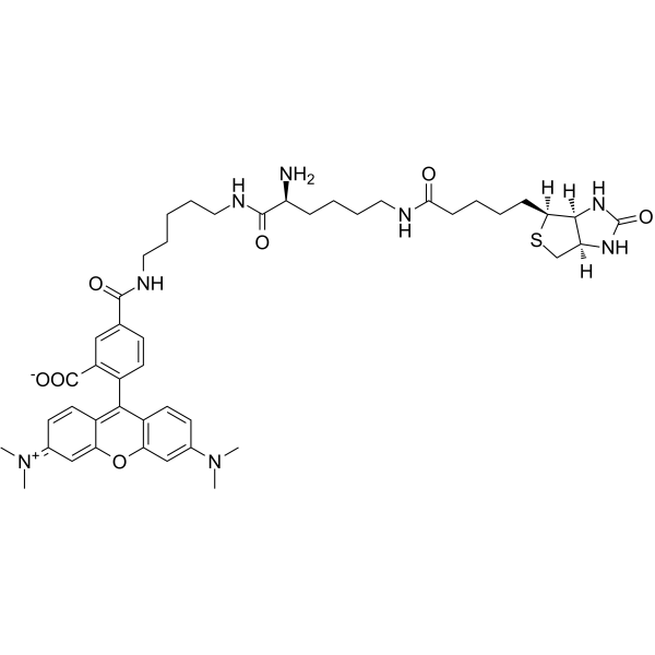
TMR Biocytin
Modify Date: 2024-04-02 18:57:05

TMR Biocytin structure
|
Common Name | TMR Biocytin | ||
|---|---|---|---|---|
| CAS Number | 749247-49-2 | Molecular Weight | 869.08 | |
| Density | N/A | Boiling Point | N/A | |
| Molecular Formula | C46H60N8O7S | Melting Point | N/A | |
| MSDS | N/A | Flash Point | N/A | |
Use of TMR BiocytinTMR Biocytin is a polar tracer used in the research of cell-cell and cell-liposome fusions, as well as membrane permeability and cellular uptake during pinocytosis. TMR Biocytin can be detected using streptavidin, and is an effective neuronal tracer in live tissue (Ex=544 nm, Em=571 nm)[1]. |
| Name | TMR Biocytin |
|---|
| Description | TMR Biocytin is a polar tracer used in the research of cell-cell and cell-liposome fusions, as well as membrane permeability and cellular uptake during pinocytosis. TMR Biocytin can be detected using streptavidin, and is an effective neuronal tracer in live tissue (Ex=544 nm, Em=571 nm)[1]. |
|---|---|
| Related Catalog | |
| In Vivo | TMR Biocytin can be used to examine the permeability changes of blood brain barrier (BBB)[2]. Guidelines (Following is our recommended protocol. This protocol only provides a guideline, and should be modified according to your specific needs)[2]. 1. Dilute 1 mg TMR Biocytin in 100 μL PBS (per mouse), and inject the solution into the tail vein. 2. 30 min after injection, anesthetize and perfuse the animals. 3. Remove spinal cords, prepare deep frozen and serial 10 μm longitudinal sections. 4. Stain nuclear counterstain using DAPI. 5. Obtaine the images of whole sections with ×10 power of objective, ×10 power of eyepiece, by using identical laser intensity, exposure times and magnification in all cohorts. 6. To set the above parameters, livers from tracer injected mice and non-injected mice were used. |
| References |
| Molecular Formula | C46H60N8O7S |
|---|---|
| Molecular Weight | 869.08 |