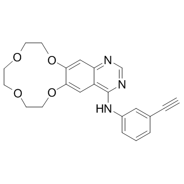
610798-31-7
| Name | Icotinib |
|---|---|
| Synonyms |
Icotinib
N-(3-Ethynylphenyl)-7,8,10,11,13,14-hexahydro[1,4,7,10]tetraoxacyclododecino[2,3-g]quinazolin-4-amine Conmana icotinib,BPI-2009H BPI-2009 [1,4,7,10]Tetraoxacyclododecino[2,3-g]quinazolin-4-amine, N-(3-ethynylphenyl)-7,8,10,11,13,14-hexahydro- UNII-9G6U5L461Q (1,4,7,10)Tetraoxacyclododecino(2,3-g)quinazolin-4-amine,N-(3-ethynylphenyl)-7,8,10,11,13,14-hexahydro |
| Description | Icotinib (BPI-2009) is a potent and specific EGFR inhibitor with an IC50 of 5 nM; also inhibits mutant EGFRL858R, EGFRL858R/T790M, EGFRT790M and EGFRL861Q. |
|---|---|
| Related Catalog | |
| Target |
EGFR:5 nM (IC50) EGFRL858R EGFRL858R/T790M EGFRT790M EGFRL861Q |
| In Vitro | Incubation with Iconitib at 0.5 μM results in kinase activity inhibition of 91%, 99%, 96%, 61% and 61%, respectively. Iconitib inhibits the proliferation of A431 and BGC-823 A549, H460 and KB cell lines with IC50s of 1, 4.06, 12.16, 16.08, 40.71 μM. When profiled with 88 kinases, Icotinib only shows meaningful inhibitory activity to EGFR and its mutants. Icotinib blocks EGFR-mediated intracellular tyrosine phosphorylation (IC50=45 nM) in the human epidermoid carcinoma A431 cell line and inhibits tumor cell proliferation[1]. |
| In Vivo | Icotinib exhibits potent dose-dependent antitumor effects in nude mice carrying a variety of human tumor-derived xenografts. The drug is well tolerated at doses up to 120 mg/kg/day in mice without mortality or significant body weight loss during the treatment. Icotinib inhibits tumor growth at a rate of 25.2%, 45.6% and 51.5% in the A431 cell line groups; 3.4%, 25.9% and 31.0% in the A549 cell line groups; 49.4%, 52.6% and 67.4% in the H460 cell line groups, and 30.3%, 36.4% and 46.5% in the HCT8 cell line groups, at 30, 60 and 120 mg/kg/dose, respectively[1]. |
| Kinase Assay | In the in vitro kinase assays, 2.4 ng/μL EGFR protein is mixed with 32 ng/μL Crk in 25 μL kinase reaction buffer containing 1 μM cold ATP and 1 μCi32P-γ-ATP. The mix is incubated with Icotinib at 0, 0.5, 2.5, 12.5 or 62.5 nM on ice for 10 min followed by incubation at 30°C for 20 min. After quenching with SDS sample bufferat 100°C for 4 min, the protein mix is resolved by electrophoresis in a 10% SDS-PAGE gel. The dried gel is then exposed to detect radioactivity. Quantification is performed by software[1]. |
| Cell Assay | Cells (1000/well) are seeded into 96-well plates in RPMI-1640 medium containing 10% FBS and grown in a 5% CO2 incubator at 37°C. After 24 h, cells are treated with Icotinib at 0, 0.78, 1.56, 3.125, 6.25, 12.5 or 25 μM for 96 h. Cell proliferation is calculated by subtracting the mean absorbance value on day 0 from the mean absorbance value on day 4[1]. |
| Animal Admin | Mice: The effect of three doses of Icotinib (30, 60, and 120 mg/kg/dose p.o. qd) on antitumor activity and survival is determined in mice bearing A431, A549, H460 and HCT8 tumor xenografts. Taxol (30 mg/kg/dose i.p. once a week) is employed in these experiments as a positive control group[1]. |
| References |
| Density | 1.3±0.1 g/cm3 |
|---|---|
| Boiling Point | 581.3±50.0 °C at 760 mmHg |
| Molecular Formula | C22H21N3O4 |
| Molecular Weight | 391.420 |
| Flash Point | 305.3±30.1 °C |
| Exact Mass | 391.153198 |
| PSA | 74.73000 |
| LogP | 1.88 |
| Vapour Pressure | 0.0±1.6 mmHg at 25°C |
| Index of Refraction | 1.649 |
