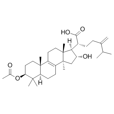
29070-92-6
| Name | Pachymic acid |
|---|---|
| Synonyms |
pachimic acid
Lanost-8-en-21-oic acid, 3-(acetyloxy)-16-hydroxy-24-methylene-, (3β,16α)- 3-O-Acetyltumulosic acid 3-ACETYLTUMULOSIC ACID Lanost-8-en-21-oic acid,3-(acetyloxy)-16-hydroxy-24-methylene-,(3beta,16alpha) (3β,16α)-3-Acetoxy-16-hydroxy-24-methylenelanost-8-en-21-oic acid Pachymasaeure (3b,16a)-3-(Acetyloxy)-16-hydroxy-24-methylenelanost-8-en-21-oic acid Lanost-8-en-21-oic acid,3-(acetyloxy)-16-hydroxy pachymaic acid |
| Description | Pachymic acid is a lanostrane-type triterpenoid from P. cocos. Pachymic acid inhibits Akt and ERK signaling pathways. |
|---|---|
| Related Catalog | |
| Target |
Akt ERK |
| In Vitro | Pachymic acid (PA) is able to inhibit gallbladder cancer tumorigenesis involving affection of Akt and ERK signaling pathways. Pachymic acid (PA) treatment significantly inhibits Rho A, Akt and ERK pathway in gallbladder carcinoma cells. Pachymic acid (PA) treatment can dose-dependently downregulate PCNA, ICAM-1, RhoA, p-Akt and pERK. Cell growth is inhibited by 10 µg/mL Pachymic acid (PA) 12 h after treatment, and a concentration of 30 µg/mL further reduced cell growth. The growth of cells is suppressed in a time- and dose-dependent manner. After 48 h treatment, about 25%, 40% and 70% of the cell growth are inhibited by Pachymic acid (PA) at concentration of 10 µg/mL, 20 µg/mL and 30 µg/mL, respectively. Pachymic acid (PA) also inhibits the growth of gallbladder carcinoma cells in a time-dependent and dose-dependent manner[1]. |
| In Vivo | To evaluate the anti-tumor activity of Pachymic acid (PA) in vivo, human lung cancer NCI-H23 tumor xenograft models are used. Pachymic acid (PA) significantly suppresses tumor growth at doses of 30 and 60 mg/kg for 21 days compared with the control group[2]. |
| Cell Assay | The antiproliferative effect of Pachymic acid (PA) on GBC-SD cells is evaluated using a cell counting kit-8 (CCK-8). Briefly, after indicated treatment, 10 µL of CCK-8 solution is added into each well, and following one hincubation, absorbance is measured at 450 nm using a microplate reader[1]. |
| Animal Admin | Mice[2] Female athymic nude mice of 4-5 weeks of age are used. Exponentially growing NCI-H23 cells (5×10⁶ in 100 µL PBS) are injected subcutaneously in the right flank of each mouse. Tumor xenografts are allowed to grow to an average size of 100-200 mm3 and are randomly assigned to four different treatment groups (six mice per group): (a) vehicle control (0.1% DMSO in physiological saline); (b) Pachymic acid (PA) 10 mg/kg; (c) PA 30 mg/kg; (Dd) PA 60 mg/kg. The mice are administered PA via intraperitoneal (ip) injections for 3 weeks (5 days/week). Tumor size is measured on two axes with the aid of Vernier calipers and tumor volume (mm3) is calculated. |
| References |
| Density | 1.1±0.1 g/cm3 |
|---|---|
| Boiling Point | 612.2±55.0 °C at 760 mmHg |
| Molecular Formula | C33H52O5 |
| Molecular Weight | 528.763 |
| Flash Point | 184.7±25.0 °C |
| Exact Mass | 528.381470 |
| PSA | 83.83000 |
| LogP | 8.59 |
| Vapour Pressure | 0.0±4.0 mmHg at 25°C |
| Index of Refraction | 1.540 |
| Storage condition | -20°C |

