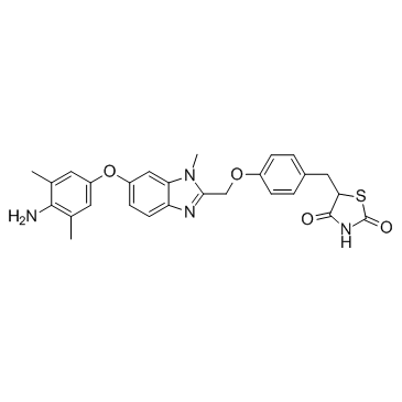
223132-37-4
| Name | 5-[[4-[[6-(4-amino-3,5-dimethylphenoxy)-1-methylbenzimidazol-2-yl]methoxy]phenyl]methyl]-1,3-thiazolidine-2,4-dione |
|---|---|
| Synonyms |
Inolitazone
Efatutazone 5-(4-{[5-(4-Amino-3,5-dimethylphenoxy)-1H-benzimidazol-2-yl]methoxy}benzyl)-1,3-thiazolidine-2,4-dione UNII-M17ILL71MC 2,4-Thiazolidinedione, 5-[[4-[[6-(4-amino-3,5-dimethylphenoxy)-1H-benzimidazol-2-yl]methoxy]phenyl]methyl]- |
| Description | Inolitazone a novel high-affinity PPARγ agonist that is dependent upon PPARγ for its biological activity with IC50 of 0.8 nM for growth inhibition. |
|---|---|
| Related Catalog | |
| Target |
PPARγ |
| In Vitro | Inolitazone (RS5444) upregulates the cell cycle kinase inhibitor, p21 WAF1/CIP1. Silencing p21 WAF1/CIP1 rendered cells insensitive to Inolitazone. A 10 nM dose of Inolitazone activates PPARγ:RXRα-dependent transcription as demonstrated in a transient transfection assay utilizing a PPRE response element fused to a luciferase reporter gene (PPRE3-tk-luc). DRO cells are treated in culture with Inolitazone, Rosiglitazone, or Troglitazone at the indicated concentrations. DRO cells are transiently transfected with PPRE3-tk-luc to examine effective concentrations at which EC50 occurs. The EC50s are 1 nM (Inolitazone), 65 nM (Rosiglitazone) and 631 nM (Troglitazone). Similarly, the calculated inhibitory concentration at IC50 is 0.8 nM for Inolitazone, 75 nM for Rosiglitazone, and 1412 nM for Troglitazone. Inolitazone specifically activates PPARγ, but not PPARα or PPARδ. Exposure of 10 nM Inolitazone following transient transfection with the appropriate PPAR isoform (γ, α, or δ) and PPAR response element linked to a luciferase reporter in RIE rat small intestinal cell line, which does not express PPARs, yields increased luciferase activity only in the presence of PPARγ and PPRE3-tk-luc[1]. DRO cells are growth inhibited by 10 nM Inolitazone (RS5444) through a PPARγ-dependent mechanism[2]. |
| In Vivo | Inolitazone (RS5444) plus Paclitaxel demonstrate additive antiproliferative activity in cell culture and minimal ATC tumor growth. When Inolitazone is administered in the diet to athymic nude mice prior to DRO tumor cell implantation, tumor growth is inhibited in a dose responsive fashion. At the highest dose, 0.025% Inolitazone inhibits growth on day 32 by 94.4% as compared to that of control. In this treatment group, five of 10 animals do not develop demonstrable tumors. In the 0.0025% treatment group, tumor growth is inhibited by 62.3% compared to that of control on day 32 while the 0.00025% dose demonstrated no growth inhibitory activity as compared to control. Tumors is nest allowed to establish in the mouse and began 0.025% Inolitazone treatment of mice 1 week after DRO or ARO tumor cell implantation. Inolitazone treated animals demonstrate tumor growth inhibition of 68.9% in DRO tumors and 48.3% in ARO tumors as compared to that of their respective controls on day 35[1]. |
| Cell Assay | DRO90-1 (DRO) and ARO81 (ARO) cells are plated in 12-well culture plates in triplicate for each condition at an initial concentration of 2×10⁴ cells/well. After overnight incubation, cells are treated with either Inolitazone, Rosiglitazone, Troglitazone, GW9662, or Paclitaxel diluted in DMSO at concentrations indicated in figure legends. All cells receive identical volumes of DMSO and are exposed to each drug for 6 days with medium and drug changed every 48 h. After 6 days, cells are washed with PBS, trypsinized and counted by Beckman Coulter Counter[1]. |
| Animal Admin | Mice[1] Suspensions of 1×10⁶/0.1 mL DRO or ARO cells in RPMI medium are injected subcutaneously in one flank of 3-4 week athymic female nude mice. Mice are changed to specialized diets either 1 week prior or 1 week after tumor implantation and randomly assigned to experimental or control groups with 10 mice per group. Diets consisted either placebo, 0.00025%, 0.0025%, or 0.025% Inolitazone formulated into the diet. Mice weighed between 20-25 g and consume on average 4 g of food per day. For combinatorial studies either placebo, 10 mg/kg or 15 mg/kg paclitaxel is injected i.p. twice weekly. Tumors are measured every 3-4 days for 35 days with calipers and tumor volumes are calculated by the formula: 0.5236(a×b×c), where a is the shortest diameter, b is the diameter perpendicular to a and c is the diameter height. |
| References |
| Density | 1.4±0.1 g/cm3 |
|---|---|
| Boiling Point | 794.6±60.0 °C at 760 mmHg |
| Molecular Formula | C27H26N4O4S |
| Molecular Weight | 502.58 |
| Flash Point | 434.4±32.9 °C |
| PSA | 144.63000 |
| LogP | 3.61 |
| Vapour Pressure | 0.0±2.8 mmHg at 25°C |
| Index of Refraction | 1.706 |
| Storage condition | 2-8℃ |
