| Description |
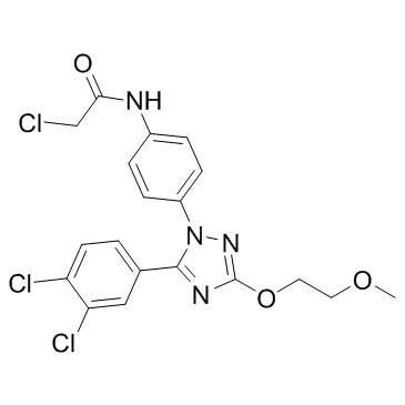
MI 2 (MALT1 inhibitor) is an irreversible MALT1 protease inhibitor with an IC50 of 5.84 μM.
|
| Related Catalog |
|
| Target |
IC50: 5.84 μM (MALT1)[1]
|
| In Vitro |
MI 2 (MI-2) is a lead compound with nanomolar activity in cell-based assays and selective activity against ABC-DLBCLs. MI 2 is the most potent in cell-based assays, with 25% growth-inhibitory concentration (GI50) values in the high-nanomolar range. MI 2 induces significant selective dose-dependent suppression of ABC-DLBCL cells (p<0.0001). HBL-1 cells are treated with increasing concentrations of MI 2 for 24 hr and cleavage of the MALT1 target protein CYLD is measured by western blotting and densitometry. MI 2 causes a dose-dependent decrease in MALT1-mediated cleavage, noted by an increase in the uncleaved CYLD protein and a decrease in the cleaved form of the protein. MI 2 is selective as a MALT1 paracaspase inhibitor, because it displays little activity against the structurally related caspase family members caspase-3, -8, and -9. Moreover, MI 2 does not inhibit caspase-3/7 activity or apoptosis in cell-based assays at concentrations that suppress MALT1[1].
|
| In Vivo |
Five C57BL/6 mice were exposed to daily intraperitoneal (IP) administration of increasing doses of MI 2 (MI-2) ranging from 0.05 to 25 mg/kg over the course of 10 days to a cumulative dose of 51.1 mg/kg, and another five mice were exposed to vehicle only (5% DMSO, n=5). There is no evidence of lethargy, weight loss, or other physical indicators of sickness. To ascertain whether the maximal administered dose of 25 mg/kg is safe in a 14 day schedule, ten mice are exposed to daily IP administration of 25 mg/kg of MI 2 over 14 days to a cumulative dose of 350 mg/kg, using as controls five mice injected with vehicle only. Five mice are sacrificed after the 14 day course of MI 2 administration (together with the five controls) and the other five mice are sacrificed after a 10 day washout period to assess delayed toxicity. No toxic effects or other indicators of sickness, including weight loss or tissue damage (macroscopic or microscopic), are noted. Brain, heart, lung, liver, kidney, bowel, spleen, thymus, and bone marrow tissues are examined. Bone marrow is normocellular with trilineage maturing hematopoiesis. Myeloid-to-erythroid ratio is 4-5:1. Megakaryocytes are normal in number and distribution. There was no fibrosis or increased number of blasts or lymphocytes. Complete peripheral blood counts, biochemistry, and liver function tests are normal[1].
|
| Cell Assay |
Cell number and viability are determined by trypan blue exclusion. Primary DLBCL cells are cultured in 96-well plates. Cells are grown in RPMI medium with 20% FBS supplemented with antibiotics for 48 hr. Cells are exposed to 0.8 µM MI 2 (1 µM for Pt.2) or control (DMSO) in quadruplicate. After 48 hr of exposure, viability is determined by using trypan blue. All samples are normalized to their own replicate control[1].
|
| Animal Admin |
Mice[1] Eight-week-old male SCID NOD.CB17-Prkdcscid/J mice are subcutaneously injected with low-passage 107 human HBL-1, TMD8, or OCI-Ly1 cells in 50% Matrigel. Treatment is initiated when tumors reach an average size of 120 mm3. C57BL/6 mice are exposed to daily intraperitoneal (IP) administration of increasing doses of MI 2 ranging from 0.05 to 25 mg/kg over the course of 10 days to a cumulative dose of 51.1 mg/kg, and another five mice are exposed to vehicle only (5% DMSO, n=5). Tumor volume is monitored by digital calipering three times a week and calculated.
|
| References |
[1]. Fontan L, et al. MALT1 small molecule inhibitors specifically suppress ABC-DLBCL in vitro and in vivo. Cancer Cell. 2012 Dec 11;22(6):812-24.
|
