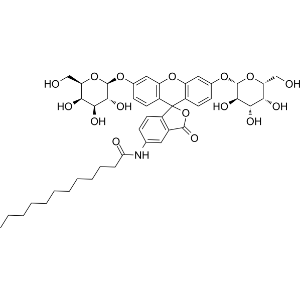
C12FDG
Modify Date: 2024-01-08 12:34:12

C12FDG structure
|
Common Name | C12FDG | ||
|---|---|---|---|---|
| CAS Number | 138777-25-0 | Molecular Weight | 853.90500 | |
| Density | N/A | Boiling Point | N/A | |
| Molecular Formula | C44H55NO16 | Melting Point | N/A | |
| MSDS | N/A | Flash Point | N/A | |
Use of C12FDGC12FDG (5-Dodecanoylaminofluorescein di-β-D-Galactopyranoside) is a lipophilic green fluorescent substrate for β-galactosidase detection. C12-FDG is more sensitive than FDG (HY-101895) for beta-galactosidase activity determinations in animal cells[1]. |
| Name | N-[3-oxo-3',6'-bis[[(2S,3R,4S,5R,6R)-3,4,5-trihydroxy-6-(hydroxymethyl)oxan-2-yl]oxy]spiro[2-benzofuran-1,9'-xanthene]-5-yl]dodecanamide |
|---|---|
| Synonym | More Synonyms |
| Description | C12FDG (5-Dodecanoylaminofluorescein di-β-D-Galactopyranoside) is a lipophilic green fluorescent substrate for β-galactosidase detection. C12-FDG is more sensitive than FDG (HY-101895) for beta-galactosidase activity determinations in animal cells[1]. |
|---|---|
| Related Catalog | |
| In Vitro | Guidelines (Following is our recommended protocol. This protocol only provides a guideline, and should be modified according to your specific needs). Fluorescent senescence-associated β-galactosidase (SA-β-Gal) assay[2]: 1. Culture cells in 6-, 12- , 24-, or 96-well plates at a density of 5× 10⁵ cells/mL overnight. Incubate the cells according to your normal protocol. 2. Wash cells by 200 μL of PBS once and fix with 100 μL of fixation solution (2% formaldehyde/0.2% glutaraldehyde in distilled water) at room temperature for 5 min. 3. Wash cells by 200 μL of PBS two times, and stain with 100 μL of 33 μM C12FDG (in PBS, pH=6.0) for 10 min, and with 200 μL of Hoechst solution (1 μg/mL Hoechst 33342 (HY-15559) in PBS, pH 6.0) for 10 min. 4. Image these cells by a 20× objective and 360-nm (Hoechst 33342) and 480-nm (C12FDG) excitation filters, and monitor through 460-nm and 535-nm emission filters, respectively. |
| References |
| Molecular Formula | C44H55NO16 |
|---|---|
| Molecular Weight | 853.90500 |
| Exact Mass | 853.35200 |
| PSA | 266.88000 |
| LogP | 3.12090 |
| Storage condition | -20°C |
| 5-Dodecanoylaminoflorescein di-b-D-galactopyranoside |
| W0468 |