| In Vitro |
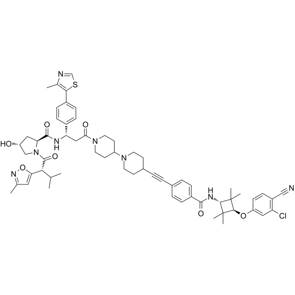
ARD-61 binds to AR protein through its AR antagonist portion and von Hippel-Lindau (VHL)/cullin 2 E3 ligase through its VHL ligand portion to recruit AR protein to cullin 2 for ubiquitination, followed by proteasome-dependent AR degradation[1]. ARD-61 (0.001-100 μM; for 7 days) has IC50 values of 235 nM and 121 nM in the MDA-MB-453 and HCC1428 cell lines, which have the highest AR expression, respectively. ARD-61 demonstrates partial cell growth inhibition, delivering IC50 values of 39, 147, and 380 nM, respectively, in the MCF-7, BT-549 and MDA-MB-415 cell lines, which have a moderate level of AR protein[1]. ARD-61 (25-100000 nM; 6-72 h) induces G2/M cell cycle arrest in a dose- and time-dependent manner in each of these three AR+ breast cancer cell lines[1]. ARD-61 (25-100000 nM; 72 h) induces apoptosis in the MDA-MB-453 and HCC1428 cell lines[1]. ARD-61 (0.01-1000 nM; 6 h) is highly potent and effective in reducing AR protein levels. ARD-61 (0.01-1000 nM; 24 h) reduces the level of PR protein with a DC50 value of 0.15 nM in the T47D cells. ARD-61 has no obvious effect on ER and GR proteins[1]. ARD-61 (1 µM; for 24 h) effectively inhibits Wnt/β-catenin and MYC signaling pathways. ARD-61 (1-1000 nM; for 24 h) not only decreases both phosphorylated HER2 and HER3, but also un-phosphorylated HER2 and HER3 proteins[1]. Efficient knock-down of VHL completely blocks AR degradation induced by ARD-61 (100 nM; 24 h) in both MDA-MB-453 and MCF-7 cell lines[1]. Cell Viability Assay[1] Cell Line: MDA-MB-453 and HCC1428 cell lines Concentration: 0.001, 0.01, 0.1, 1, 10, 100 μM Incubation Time: 7 days Result: Achieves near complete inhibition of cell growth. Cell Cycle Analysis[1] Cell Line: MDA-MB-453, HCC1428 and MCF-7 cell lines Concentration: 25, 250, 500, 1000, 10000, 100000 nM Incubation Time: 6-72 hours Result: Induced G2/M cell cycle arrest in a dose- and time-dependent manner in each of these three AR+ breast cancer cell lines. Apoptosis Analysis[1] Cell Line: MDA-MB-453 and HCC1428 cell lines Concentration: 25, 250, 500, 1000, 10000, 100000 nM Incubation Time: 6-72 hours Result: Induced apoptosis in the MDA-MB-453 and HCC1428 cell lines in a dose-dependent manner. Western Blot Analysis[1] Cell Line: MDA-MB-453, MCF-7, BT549, MDA-MB-415 and HCC1428 cell lines Concentration: 0.01, 0.03, 0.1, 0.3, 1, 3, 10, 30, 100, 300, 1000 nM Incubation Time: 6 hours Result: Reduced AR protein levels in the MDA-MB-453 (DC50=0.44 nM), MCF-7 (DC50=1.8 nM), BT549 (DC50=2.0 nM), MDA-MB-415 (DC50=2.4 nM) and HCC1428 (DC50=3.0 nM) cell lines.
|
