| In Vitro |
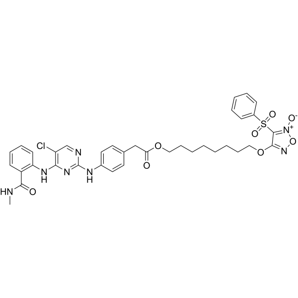
FAK-IN-9 (Compound 8f; 72 h) 抑制 MDA-MB-157、MDA-MB-231 和 MDA-MB-453 细胞增殖,IC50 分别为 0.167±0.025、0.126±0.012 和 0.159±0.017 μM [1]。 FAK-IN-9 (1-4 μM; 72 h) 导致 MDA-MB-231 细胞以剂量依赖的方式产生相对较高水平的 NO[1]。 FAK-IN-9 (1-4 μM; 48 h) 抑制 MDA-MB-231 细胞的侵袭和迁移[1]。 FAK-IN-9 (1-4 μM; 72 h) 有效阻断 FAK 介导的信号通路[1]。 FAK-IN-9 (4 μM; 72 h) 抑制 MDA-MB-231 细胞局灶粘连 (FAs) 和应力纤维 (SFs) 的形成[1]。 FAK-IN-9 (1-4 μM; 72 h) 诱导 MDA-MB-231 细胞凋亡[1]。 Cell Proliferation Assay[1] Cell Line: MDA-MB-157, MDA-MB-231, MDA-MB-453 and MCF10A Concentration: Incubation Time: 72 h Result: Inhibited proliferation with IC50s of 0.167±0.025, 0.126±0.012, 0.159±0.017 and 2.401±0.131 μM against MDA-MB-157, MDA-MB-231, MDA-MB-453 and MCF10A, respectively. Cell Invasion Assay[1] Cell Line: MDA-MB-231 cells Concentration: 1, 2 and 4 μM Incubation Time: 48 h Result: The numbers of invasive MDA-MB-231 cells were reduced dose-dependently. Cell Migration Assay [1] Cell Line: MDA-MB-231 cells Concentration: 1, 2 and 4 μM Incubation Time: 48 h Result: Remarkably block the migration of MDA-MB-231 cells in a dose-dependent manner. Western Blot Analysis[1] Cell Line: MDA-MB-231 cells Concentration: 1, 2 and 4 μM Incubation Time: 72 h Result: Potently suppressed the autophosphorylation of Y397 in a dose-dependent manner. Decreased the levels of p-AKT, MMP-2 and MMP-9 dose dependently. Apoptosis Analysis[1] Cell Line: MDA-MB-231 cells Concentration: 1, 2 and 4 μM Incubation Time: 72 h Result: The percentage of apoptotic MDA-MB-231 cells gradually increased ranging from 19.06% to 77.66% at 4 μM.
|
