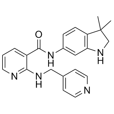
Motesanib
Modify Date: 2024-01-02 17:28:09

Motesanib structure
|
Common Name | Motesanib | ||
|---|---|---|---|---|
| CAS Number | 453562-69-1 | Molecular Weight | 373.451 | |
| Density | 1.3±0.1 g/cm3 | Boiling Point | 517.3±50.0 °C at 760 mmHg | |
| Molecular Formula | C22H23N5O | Melting Point | N/A | |
| MSDS | N/A | Flash Point | 266.6±30.1 °C | |
Use of MotesanibMotesanib is a potent ATP-competitive inhibitor of VEGFR1/2/3 with IC50s of 2 nM/3 nM/6 nM, respectively, and has similar activity against Kit, and is appr 10-fold more selective for VEGFR than PDGFR and Ret. |
| Name | motesanib |
|---|---|
| Synonym | More Synonyms |
| Description | Motesanib is a potent ATP-competitive inhibitor of VEGFR1/2/3 with IC50s of 2 nM/3 nM/6 nM, respectively, and has similar activity against Kit, and is appr 10-fold more selective for VEGFR than PDGFR and Ret. |
|---|---|
| Related Catalog | |
| Target |
VEGFR1:2 nM (IC50) VEGFR2:3 nM (IC50) VEGFR3:6 nM (IC50) |
| In Vitro | Motesanib has broad activity against the human VEGFR family, and displays > 1000 selectivity against EGFR, Src, and p38 kinase. Motesanib significantly inhibits VEGF-induced cellular proliferation of HUVECs with an IC50 of 10 nM, while displaying little effect at bFGF-induced proliferation with an IC50 of >3,000 nM. Motesanib also potently inhibits PDGF-induced proliferation and SCF-induced c-kit phosphorylation with IC50 of 207 nM and 37 nM, respectively, but not effective against the EGF-induced EGFR phosphorylation and cell viability of A431 cells[1]. Althouth displaying little antiproliferative activity on cell growth of HUVECs alone, Motesanib treatment significantly sensitizes the cells to fractionated radiation[2]. |
| In Vivo | Motesanib (100 mg/kg) significantly inhibits VEGF-induced vascular permeability in a time-dependent manner. Oral administration of Motesanib twice daily or once daily potently inhibits, in a dose-dependent manner, VEGF-induced angiogenesis using the rat corneal model with ED50 of 2.1 mg/kg and 4.9 mg/kg, respectively. Motesanib induces a dose-dependent tumor regression of established A431 xenografts by selectively targeting neovascularization in tumor cells[1]. Motesanib in combination with radiation displays significant anti-tumor activity in head and neck squamous cell carcinoma (HNSCC) xenograft models[2]. Motesanib treatment also induces significant dose-dependent reductions in tumor growth and blood vessel density of MCF-7, MDA-MB-231, or Cal-51 xenografts, which can be markedly enhanced when combined with docetaxel or tamoxifen[3]. |
| Kinase Assay | Optimal enzyme, ATP, and substrate (gastrin peptide) concentrations are established for each enzyme using homogeneous time-resolved fluorescence (HTRF) assays. Motesanib is tested in a 10-point dose-response curve for each enzyme using an ATP concentration of two-thirds Km for each. Most assays consist of enzyme mixed with kinase reaction buffer [20 mM Tris-HCl (pH 7.5), 10 mM MgCl2, 5 mM MnCl2, 100 mM NaCl, 1.5 mM EGTA]. A final concentration of 1 mM DTT, 0.2 mM NaVO4, and 20 μg/mL BSA is added before each assay. For all assays, 5.75 mg/mL streptavidin-allophycocyanin and 0.1125 nM Eu-PT66 are added immediately before the HTRF reaction. Plates are incubated for 30 minutes at room temperature and read on a Discovery instrument. IC50 values are calculated using the Levenberg-Marquardt algorithm into a four-parameter logistic equation. |
| Cell Assay | Cells are preincubated for 2 hours with different concentrations of Motesanib, and exposed with 50 ng/mL VEGF or 20 ng/mL bFGF for an additional 72 hours. Cells are washed twice with DPBS, and plates are frozen at -70°C for 24 hours. Proliferation is assessed by the addition of CyQuant dye, and plates are read on a Victor 1420 workstation. IC50 data are calculated using the Levenberg-Marquardt algorithm into a four-parameter logistic equatio. |
| Animal Admin | A431 cells are cultured in DMEM (low glucose) with 10% FBS and penicillin/streptomycin/glutamine. Cells are harvested by trypsinization, washed, and adjusted to a concentration of 5×10⁷/mL in serum-free medium. Animals are challenged s.c. with 1×10⁷ cells in 0.2 mL over the left flank. Approximately 10 days thereafter, mice are randomized based on initial tumor volume measurements and treated with either vehicle (Ora-Plus) or Motesanib. Tumor volumes and body weights are recorded twice weekly and/or on the day of sacrifice. Tumor volume is measured with a Pro-Max electronic digital caliper and calculated using the formula length (mm)×width (mm)×height (mm) and expressed in mm3. Data are expressed as mean±SE. Repeated measures ANOVA followed by Scheffe post hoc testing for multiple comparisons is used to evaluate the statistical significance of observed differences. |
| References |
| Density | 1.3±0.1 g/cm3 |
|---|---|
| Boiling Point | 517.3±50.0 °C at 760 mmHg |
| Molecular Formula | C22H23N5O |
| Molecular Weight | 373.451 |
| Flash Point | 266.6±30.1 °C |
| Exact Mass | 373.190247 |
| PSA | 78.94000 |
| LogP | 4.22 |
| Vapour Pressure | 0.0±1.3 mmHg at 25°C |
| Index of Refraction | 1.669 |
| Storage condition | -20°C |
| Motesanib |
| N-(3,3-Dimethyl-2,3-dihydro-1H-indol-6-yl)-2-[(pyridin-4-ylmethyl)amino]nicotinamide |
| N-(3,3-Dimethyl-2,3-dihydro-1H-indol-6-yl)-2-[(4-pyridinylmethyl)amino]nicotinamide |
| 3-pyridinecarboxamide, N-(2,3-dihydro-3,3-dimethyl-1H-indol-6-yl)-2-[(4-pyridinylmethyl)amino]- |
| N-(3,3-dimethylindolin-6-yl)-2-(pyridin-4-ylmethylamino)nicotinamide |
| Motesanib Base |