| In Vivo |
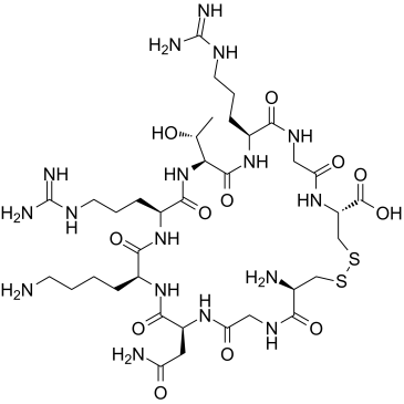
LyP-1 (8 weeks) shows a remarkable reduction in plaque formation and plaque occupation rates in the LyP-1-treated group. In addition, a higher apoptotic rate in macrophages released from hypoxic plaques is observed after the treatment of LyP-1when compared to control peptide[1]. The LyP-1 peptide is labeled with a near-infrared fluorophore (Cy5.5) for optical imaging. At days 3, 7, 14 and 21 after inoculation with 4T1 cells, tumor-bearing BALB/C mice is injected Cy5.5-LyP-1 (0.8 nmol) through the middle phalanges of the upper extremities of the tumor-bearing mice. The fluorescence intensities were 0.024, 0.038, 0.048 and 0.106 × 10⁶ photon/cm2/sec respectively at days 3, 7, 14 and 21 after tumor cell inoculation, which are 1.02, 1.63, 2.04, and 4.52-fold higher than in the contralateral LNs. Cy5.5-LyP-1 staining in LNs co-localized with LYVE-1, suggesting lymphatics-specific binding of LyP-1 peptide[1].
|


