| Description |
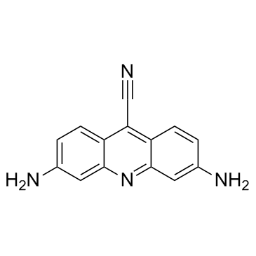
CTX1 is a novel small molecule p53 activator.
|
| Related Catalog |
|
| Target |
p53[1]
|
| In Vitro |
CTX1 induces significant p53-dependent cell death preferentially in HdmX-expressing cells as compare to cells in which p53 is inactivated by Hdm2 overexpression or p53 is knocked down using p53 targeting shRNA. CTX1 can induce p53 protein levels in the p53 mutant cell line, HT29. It is also found that CTX1 exhibits potent activity (LD50 ~1 μM) as a single agent on primary AML patient samples in a similar fashion to AML cell lines. After 16 hr of treatment with CTX1 in HCT116 p53-wild-type cells there is a decrease in S phase from 23% to 3% while HCT116 p53-null cells exhibit a reduction in S phase from 34% to 28%. The combination of low doses of CTX1 and nutlin-3 leads to a significant enhancement of apoptosis in p53-wildtype, but not p53-null cells[1].
|
| In Vivo |
CTX1 even as a single agent significantly enhances the survival of mice in this model system. Mice treated with CTX1 alone or in combination with nutlin-3 has a significantly increased survival time (p<0.0001)[1].
|
| Kinase Assay |
Exponentially growing OCI cells cultures are treated with 3 μM CTX1, 8 μM Nutlin and or 15 μM RO-5963 for 4.5 hrs. Whole-cell extracts are generated using modified RIPA lysis buffer 25 mM Tris (pH 8.0), 100 mM NaCl, 0.5 mM EDTA, 0.50% NP-40 and complete protease mixture tablet. Protein extracts (~750 μg) are precleared and immune precipitation is performed using a kit according to the manufacturer's protocol. For the immunoprecipitation, mouse monoclonal anti-p53 and rabbit polyclonal anti-HDMX are used. Immune complexes are then collected, proteins are eluted and subjected to Western blotting with the indicated antibodies[1].
|
| Animal Admin |
6 week old female NOD/SCID/IL2Rγ−/− mice are injected into the tail vein with 5×10⁶ cells primary human AML cells (n=5 per group). Drug treatment is started 2 days after tumor cell injection. Nutlin-3 is given by oral gavage (200 mg/kg) and CTX1 is injected i.p. (30 mg/kg) five days a week for 3 weeks. Flow cytometry is performed on bone marrow cells isolated from the mouse femur using a human specific CD45PE antibody using a cytometer to confirm AML infiltration into the bone marrow[1].
|
| References |
[1]. Karan G, et al. Identification of a Small Molecule That Overcomes HdmX-Mediated Suppression of p53. Mol Cancer Ther. 2016 Apr;15(4):574-582.
|
