| In Vitro |
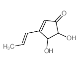
Treatment of Mel-Ab cells with Terrein (10-100 µM) for 4 days significantly reduces melanin levels in a dose-dependent manner. Terrein reduces melanin synthesis by reducing tyrosinase production via ERK activation[1]. Terrein (5-500 µM; 24 and 48 hours) reduces the viability of these cells in concentration- and time-dependent manners[2]. Terrein (150, 250, 500µM; 24 hours) induces programmed cell death in both invasive and non-invasive breast cancer cell lines. Thus, the optimal non-toxic concentrations of Terrein (25 and 75 µM) were used for subsequent experiments[2]. Terrein (25 and 75 µM) significantly decrease the mRNA levels of both MMP-2 and MMP-9 in MDA-MB-231 cells. Terrein (75 µM) significantly decrease the mRNA levels of both MMP-2 and MMP-9 in MCF-7 cells[2]. Terrein (75 µM) inhibits the expression of RhoA, RhoB, p-Rac1 in MCF-7 cells. Terrein (75 µM) inhibits the expression of RhoB, p-Rac1 in MDA-MB-231 cells[2]. Terrein (10, 30, and 100 μM) inhibits the production of virulence factors such as elastase, pyocyanin, and rhamnolipid, as well as biofilm formation in P. aeruginosa PAO1 and PA14 strains[3]. Cell Viability Assay[2] Cell Line: MCF-7 and MDA-MB-231 cells Concentration: 5, 25, 50, 75, 100, 150, 250, 500 µM Incubation Time: 24 and 48 hours Result: The IC50s for MCF-7 and MDA-MB-231 cells after 24 h of incubation were 2.34 mM and 700 µM, respectively. After 48 h of incubation, the IC50s are 244.3 µM for MCF-7 and 244.5 µM for MDA-MB-231 cells. Apoptosis Analysis[2] Cell Line: MCF-7 and MDA-MB-231 cells Concentration: 25, 75, 150, 250, 500 µM Incubation Time: 24 hours Result: The percentages of apoptotic cells after 25 and 75 µM treatment were 7.52 and 5.82%, respectively for MCF-7 cells (9.94% for the control), and 15.77 and 15.82%, respectively for MDA-MB-231 cells (8.79% for the control). RT-PCR[2] Cell Line: MCF-7 and MDA-MB-231 cells Concentration: 25, 75 µM Incubation Time: Result: Decreased the mRNA levels of both MMP-2 and MMP-9 in a concentration-dependent manner. Western Blot Analysis[2] Cell Line: MCF-7 and MDA-MB-231 cells Concentration: 75 µM Incubation Time: 6, 12 and 24 hours Result: RhoA and RhoB protein levels were significantly decreased after 6 h of 75 µM treatment in MCF-7 cells. Phosphorylated (p)-Rac1 was also slightly decreased at 12 h. However, RhoC and Cdc42 levels remained unchanged. RhoB protein levels and Rac1 phosphorylation were also decreased in MDA-MB-231 cells at 12 and 24 h, respectively.
|

 CAS#:64-18-6