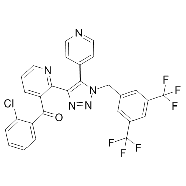
| Description |
Tradipitant is a neurokinin-1 (NK-1) antagonist.
|
| Related Catalog |
|
| Target |
NK-1[1]
|
| In Vitro |
Results reveal that Tradipitant ([3H]-LY686017) bind to sections of guinea pig brain with a distribution similar to that previously reported for NK-1 receptors. Areas with the highest density are the accessory olfactory nucleus, anteroventral thalamic nucleus, paraventricular hypothalamic nucleus central amygdaloid nucleus (lateral) and the medial amygdaloid nucleus (anterodorsal)[1].
|
| In Vivo |
Autoradiography experiments reveal that intravenously administered Tradipitant ([3H]-LY686017) localizes in the guinea pig brain and has a distribution similar to that described for the NK-1 receptor using agonist or antagonist radioligands[1].
|
| Cell Assay |
Brains from male, Hartley guinea pigs (350 to 400 g) are removed, rinsed for 5 to 15 min in cold PBS, patted dry with a paper towel and frozen on aluminum foil lined with parafilm placed on dry ice. Once frozen, the brains are stored at -80°C until they are sectioned using a cryostat set at a thickness of 12 μm. The tissue sections are thaw mounted onto chrome alum/gelatin coated slides and allowed to air dry for 5 to 20 min before being frozen again. To assessthe binding of Tradipitant ([3H]-LY686017) to sections, the slides are preincubated for 30 min in modified Kreb’s phosphate buffer followed by an incubation in the same buffer containing 0.05% bacitracin, 0.4% BSA and various concentrations of Tradipitant. Rinse time, incubation time and saturation binding are determined using sections through the caudate nucleus[1].
|
| Animal Admin |
Male, Hartley guinea pigs (550 to 650 g) are used. Guinea pigs are singly housed in cages for 2 to 4 days before being used in an experiment. On the day of the experiment, the catheters are flushed with heparinized 0.9% saline and, if the catheter is clear, the animal received 25 microCi of Tradipitant ([3H]-LY686017) in saline for time course and compound displacement studies (n=4 to 6/group) and 100 microCi for autoradiography studies (n=5). The catheter line is cleared of radioactivity by again flushing the line with 0.5 mL heparinized saline. The end of the catheter is subsequently plugged using the original steel wire plug. Based on data from initial time course experiments, the animals are sacrificed by rapid decapitation 2 h post-jugular injection for compound displacement and autoradiography studies[1].
|
| References |
[1]. Gackenheimer SL, et al. In vitro and ex vivo autoradiography of the NK-1 antagonist [³H]-LY686017 in Guinea pig brain. Neuropeptides. 2011 Apr;45(2):157-64.
|
