| Description |
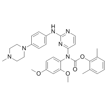
WH-4-023 is a potent and selective dual Lck/Src inhibitor with IC50 of 2 nM/6 nM for Lck and Src kinase respectively; little inhibition on p38α and KDR.
|
| Related Catalog |
|
| Target |
IC50: 2 nM (Lck), 6 nM (Src)[1]
|
| In Vitro |
WH-4-023 shows a similar potency increase on Lck as 2-substituted variants[1].
|
| Kinase Assay |
The Lck HTRF kinase assay involves ATP-dependent phosphorylation of a biotinylated substrate peptide of gastrin in the presence or absence of inhibitor compound. The final concentration of gastrin is 1.2 μM. The final concentration of ATP is 0.5 μM (Km app =0.6±0.1 μM), and the final concentration of Lck (a GST-kinase domain fusion (AA 225−509)) is 250 pM. Buffer conditions are as follows: 50 mM HEPES pH=7.5, 50 mM NaCl, 20 mM MgCl2, 5 mM MnCl2, 2 mM DTT, 0.05% BSA. The assay is quenched and stopped with 160 μL of detection reagent. Detection reagents are as follows: Buffer made of 50 mM Tris, pH=7.5, 100 mM NaCl, 3 mM EDTA, 0.05% BSA, 0.1% Tween20. Prior to reading, Streptavidin allophycocyanin (SA-APC) is added at a final concentration in the assay of 0.0004 mg/mL, along with europilated anti-phosphotyrosine Ab (Eu-anti-PY) at a final conc of 0.025 nM. The assay plate is read in a Discovery fluorescence plate reader with excitation at 320 nm and emission at 615 and 655 nm[1].
|
| Cell Assay |
The purpose of this assay is to test the potency of T cell receptor (TCR; CD3) and CD28 signaling pathway inhibitors in human T cells. T cells are purified from human peripheral blood lymphocytes (hPBL) and preincubated with or without compound prior to stimulation with a combination of an anti-CD3 and an anti-CD28 antibody in 96-well tissue culture plates (1×10⁵ T cells/well). Cells are cultured for ~20 h at 37°C in 5% CO2 and then secreted IL-2 in the supernatants is quantified by cytokine ELISA. The cells remaining in the wells are then pulsed with 3H-thymidine overnight to assess the T cell proliferative response. Cells are harvested onto glass fiber filters and 3H-thymidine incorporation into DNA is analyzed by liquid scintillation counter. For comparison purposes, phorbol myristic acid (PMA) and calcium ionophore are used in combination to induce IL-2 secretion from purified T cells. Potential inhibitor compounds are tested for inhibition of this response as described above for anti-CD3 and -CD28 antibodies. Human whole-blood anti-CD3/CD28-induced IL-2 secretion assays are run in a similar fashion as described above using whole blood from normal volunteers diluted 50% in tissue culture medium prior to stimulation[1].
|
| References |
[1]. Martin MW, et al. Novel 2-aminopyrimidine carbamates as potent and orally active inhibitors of Lck: synthesis, SAR, and in vivo antiinflammatory activity. J Med Chem. 2006 Aug 10;49(16):4981-91.
|


