| Description |
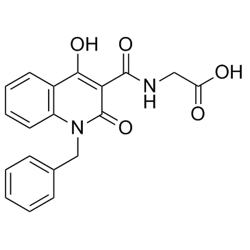
IOX2 is a specific prolyl hydroxylase-2 (PHD2) inhibitor with IC50 of 22 nM.
|
| Related Catalog |
|
| Target |
IC50: 22 nM (PHD2)[1]
|
| In Vitro |
IOX2 is at least 2-5000 fold selective, as judged by IC50 values, for PHD2 over the KDMs and Factor Inhibiting HIF(FIH)[1]. IOX2 significantly upregulates the transcription of VEGF-A and BNIP3 in normal human epidermal keratinocytes (NHEK) and normal human dermal fibroblasts (NHDF) when grown under normoxia and hypoxia. IOX2 efficiently promotes HIF-1α stability, nuclear translocation, and target gene expression in keratinocytes and fibroblasts. In addition, IOX2 significantly upregulates biosynthesis and transcription of VEGF-A and BNIP3 in sulfur mustard (SM)-exposed NHEK and NHDF grown under hypoxia. These results suggest that application of IOX2 is useful for restoring of SM-affected HIF-1α stability and signaling activity in keratinocytes and fibroblasts[2].
|
| In Vivo |
To investigate the utility of IOX2 as in vivo functional probes, IOX2 is tested to upregulate HIF signaling in a whole organism, that is, transgenic zebrafish (Danio rerio). Because the expression of the PHD3 encoding gene is regulated by HIF in humans and zebrafish, PHD3 levels are a readout of HIF activity. A zebrafish hypoxia reporter line is generated expressing GFP with the phd3 promoter elements. Transgenic wild-type embryos at 3 days postfertilization treated with compounds (10 μM) for 2 days displayed clear increase in phd3:EGFP expression in the liver, relative to controls. Significant increases in GFP levels are observed with IOX2[1].
|
| Kinase Assay |
Inhibition assays are carried out in 384-well white ProxiPlates in 10 μL of reaction volume. Standard reaction mixtures consisted of the compound (in 2% DMSO final concentration), enzyme mix (0.001 μM of PHD2, 10 μM of Fe(II), 100 μM of ascorbate) and peptide mix (0.06 μM of biotinylated C-terminal oxygen dependent degradation domain (CODD) peptide, 2 μM of 2OG) in 50 mM HEPES pH 7.5, 0.01% Tween-20 and 0.1% BSA buffer. Compounds (e.g., IOX2) are preincubated with the enzyme mix for 15 min before being incubated with peptide mix for 10 min at 22°C. Each reaction is quenched with 5 μL of 30 mM EDTA. The bead mix containing AlphaScreen beads is preincubated for 1h with a rabbit monoclonal antibody selective for hydroxy-HIF1α (Pro564) and are added to the wells for a further 1 h at 22°C. The plates are then analyzed with an Envision plate reader. The IC50 values are calculated using nonlinear regression with normalized dose-response fit using Prism GraphPad (n≥3)[1].
|
| Cell Assay |
Both VHL-defective (renal carcinoma cells with an empty vector, RCC4) and VHL-competent cells human embryonic kidney HEK293T, osteosarcoma U2OS and RCC4/VHLHA (RCC4 stably transfected with C-terminal HA-tagged wt VHL) are used. Cells are treated with DMSO (control) and tested compounds (e.g., IOX2) (dissolved in DMSO except for DMOG which is dissolved in PBS and added directly to culture medium) for 4-5 h. Cell extracts are probed with antibodies to hydroxy-Pro564 (CODD-OH) and hydroxy-Asn803 (CAD-OH). HIF-1α band intensities are used to normalize hydroxylation signals[1].
|
| Animal Admin |
Zebrafish[1] Phd3:gfpsh144/sh144 fish (Danio rerio) are incrossed to produce phd3:gfpsh144/sh144 embryos, these are raised at 28°C in E3 medium. The phd3:gfpsh144/sh144 line is a hypoxia reporter line created by BAC recombination of the phd3 reporter GFP construct. At 3 days post fertilization, potential inhibitors (e.g., IOX2) are added in fresh medium at 10 μM in 1% DMSO. The embryos are incubated with the compounds for a further 48 h. Embryos are anesthetized at 5 dpf by immersion in tricaine. Lateral view images of the embryos are taken using a fluorescent dissecting stereomicroscope (both bright-field and fluorescent). Fluorescent images are analyzed using Image J.
|
| References |
[1]. Chowdhury R, et al. Selective small molecule probes for the hypoxia inducible factor (HIF) prolyl hydroxylases. ACS Chem Biol. 2013 Jul 19;8(7):1488-96. [2]. Deppe J, et al. Impairment of hypoxia-induced HIF-1α signaling in keratinocytes and fibroblasts by sulfur mustard is counteracted by a selective PHD-2 inhibitor. Arch Toxicol. 2016 May;90(5):1141-50.
|

