EGFR-IN-60
Modify Date: 2024-01-13 20:36:38
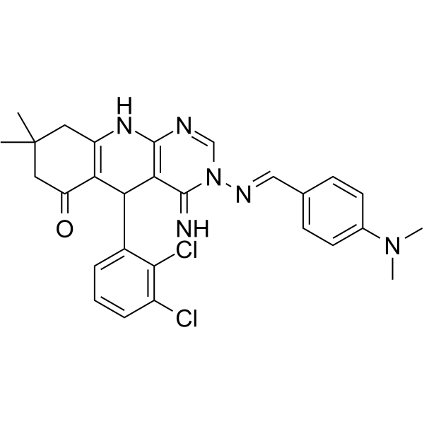
EGFR-IN-60 structure
|
Common Name | EGFR-IN-60 | ||
|---|---|---|---|---|
| CAS Number | 2699877-43-3 | Molecular Weight | 535.47 | |
| Density | N/A | Boiling Point | N/A | |
| Molecular Formula | C28H28Cl2N6O | Melting Point | N/A | |
| MSDS | N/A | Flash Point | N/A | |
Use of EGFR-IN-60EGFR-IN-60 (Compound 7d) shows obvious inhibition of EGFRWT, EGFRT790M, EGFRL858R and JAK3 with IC50s of 83, 26, 53, and 69 nM, respectively. EGFR-IN-60 potently inhibits the growth of H1975 cells harboring EGFRT790M mutation (IC50=1.32 µM) over A431 cells overexpressing EGFRWT (IC50=4.96 µM). EGFR-IN-60 exhibits good oral absorption, potent and safe antitumor activity. EGFR-IN-60 induces cell death through apoptosis supported by increased Bax/Bcl-2 ratio[1]. |
| Name | EGFR-IN-60 |
|---|
| Description | EGFR-IN-60 (Compound 7d) shows obvious inhibition of EGFRWT, EGFRT790M, EGFRL858R and JAK3 with IC50s of 83, 26, 53, and 69 nM, respectively. EGFR-IN-60 potently inhibits the growth of H1975 cells harboring EGFRT790M mutation (IC50=1.32 µM) over A431 cells overexpressing EGFRWT (IC50=4.96 µM). EGFR-IN-60 exhibits good oral absorption, potent and safe antitumor activity. EGFR-IN-60 induces cell death through apoptosis supported by increased Bax/Bcl-2 ratio[1]. |
|---|---|
| Related Catalog | |
| Target |
EGFR:0.083 μM (IC50) EGFRL858R:0.053 μM (IC50) EGFRT790M:0.026 μM (IC50) JAK3:0.069 μM (IC50) |
| In Vitro | EGFR-IN-60 (compound 7d) (3.25-88.46 μM, 48 hours) shows well antitumor activity against hepatocellular (HepG2), colorectal (HCT-116) and breast (MCF-7) cancer cells[1]. EGFR-IN-60 (compound 7d) (0.49-86.4 μM, 48 hours) shows cytotoxic activity against cancer cells[1]. EGFR-IN-60 (compound 7d) (0-5.27 μM, 24 hours) induces an increase in G2/M phase cells and induces apoptosis in HepG2, HCT-116, and MCF-7 cell lines[1]. EGFR-IN-60 (compound 7d) (0 µM, 10 µM, 24 hours) can induce apoptosis through up-regulation of Bax and down-regulation of Bcl-2[1]. Cell Proliferation Assay[1] Cell Line: Hepatocellular (HepG2), Colorectal (HCT-116), Breast (MCF-7) cancer cells Concentration: 3.25-88.46 μM Incubation Time: 48 hours Result: Inhibited HepG2 cells, HCT-116 cells, MCF-7 cells with IC50values of 4.46 μM, 5.27 μM and 3.25 μM respectively. Cell Cytotoxicity Assay[1] Cell Line: Overexpress EGFRWT human epidermoid carcinoma cells (A431), Mutant EGFRT790M cells NSCLC (H1975), Lung fibroblast cells (WI38), Amnion epithelial cells (WISH) Concentration: 0.49-86.4 μM Incubation Time: 48 hours Result: Showed cytotoxic activity against A431, H1975, WI38, WISH with IC50 value of 4.96 μM, 1.32 μM, 64.27 μM, 46.38 μM respectively. Cell Cycle Analysis[1] Cell Line: HepG2, HCT-116, MCF-7 Concentration: 0 μM, 3.25 μM, 4.46 μM, 5.27 μM Incubation Time: 24 hours Result: Resulted in an increase in the percentage of G2/M phase cells from 14.09% to 25.66% , from 15.87% to 38.51%, from 10.95% to 41.60% in HepG2, HCT-116, MCF-7 cell lines respectively. Apoptosis Analysis[1] Cell Line: HepG2, HCT-116, MCF-7 Concentration: 3.25 μM, 4.46 μM, 5.27 μM Incubation Time: 24 hours Result: Induced more apoptosis in MCF-7 cells comparing with HepG2 and HCT-116 cells. Western Blot Analysis[1] Cell Line: HepG2, HCT-116, MCF-7 Concentration: 0 µM, 10 µM Incubation Time: 24 hours Result: Showed the levels of pro-apoptotic protein Bax upgrading by 5.71, 8.15 and 16.51 fold and the levels of anti-apoptotic protein Bcl-2 down-regulating by 0.72, 0.53 and 0.31 fold in HepG2, HCT-116, MCF-7, respectively. |
| References |
| Molecular Formula | C28H28Cl2N6O |
|---|---|
| Molecular Weight | 535.47 |