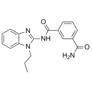
1111556-37-6
| Name | Takinib |
|---|
| Description | Takinib is a potent and selective TAK1 inhibitor with an IC50 of 9.5 nM. |
|---|---|
| Related Catalog | |
| Target |
TAK1:9.5 nM (IC50) IRAK4:120 nM (IC50) IRAK1:390 nM (IC50) GCK:430 nM (IC50) CLK2:430 nM (IC50) MINK1:1.9 μM (IC50) |
| In Vitro | At 10 mM, Takinib shows significant inhibitory activity (<10% enzyme activity after exposure) on six serine/threonine kinases, including TAK1, IRAK4, IRAK1, GCK, CLK2, and MINK1. Analysis reveals that increasing concentrations of Takinib leads to a decrease in Vmax while maintaining KM. When the enzyme is activated with 5 mM ATP for 3 hr, the same Vmax is reached for 0, 10, 50, and 100 nM Takinib, and KM increases for these concentrations, which implies that Takinib is an ATP-competitive inhibitor if TAK1 is ATP activated. Importantly, results show that Takinib inhibits the function of both activated and un-activated TAK1 with identical potency. TNF-α stimulation in the presence of Takinib induces caspase activity in MDA-MB-231 cells in a dose-dependent manner, whereas unstimulated cells do not upregulate caspase activity. Takinib reduces phosphorylation significantly but does not influence total protein levels. Takinib inhibits phosphorylation of IKK, MAPK 8/9, and c-Jun in a dose-dependent manner. Takinib shows an almost complete inhibition of IL-6 secretion at micromolar concentrations following 24 hr of treatment in the presence of TNF-α[1]. |
| Kinase Assay | Activity of purified TAK1-TAB1 protein is measured. In brief, TAK1-TAB1 (50 ng/well) is incubated with 5 μM ATP containing radiolabeled [32P]-ATP in the presence of 300 mM substrate peptide (RLGRDKYKTLRQIRQ) in a final volume of 40 μL in the presence of buffer (containing 50 mM Tris pH 7.5, 0.1 mM EGTA, 0.1% β-Mercaptoethanol, 10 mM magnesium acetate, 0.5 mM MnCl) and indicated compounds. The reaction is let go for 10 min and stopped with 10 μL concentrated H3PO4. The remaining activity is measured using a scintillation counter. Dose-response curves are repeated 3 times. For kinetic mechanistic studies, experiments are repeated two times and averaged[1]. |
| Cell Assay | MDA-MB-231 cells (1,000 cells/well) are seeded in a 96-well plate with 10% FBS, 5% Pen/Strep, 4 g/L glucose DMEM medium. After 24h, cells are serum starved with 1% FBS, 5% Pen/Strep, 4 g/L glucose DMEM medium for 4h. Cells are treated with titrations of Takinib in the presence or absence of 30 ng/mL TNFα. Plates at 0 h and 24 h following treatment are frozen at -80°C after removal of media. After 24 h, 100 μL ddH2O is added to each well and plates are refrozen. 1 μL from Hoechst stock [1 mg/mL in 1:4 DMSO/H2O] is dissolved in 1 mL of TNE buffer (10 mM Tris, 2 M NaCl, 1 mM Na2EDTA) and 100 μL of this solution is added to each well. The fluorescence is determined at 355/460 nm[1]. |
| References |
| Molecular Formula | C18H18N4O2 |
|---|---|
| Molecular Weight | 322.36 |
| Storage condition | -20℃ |
