| Description |
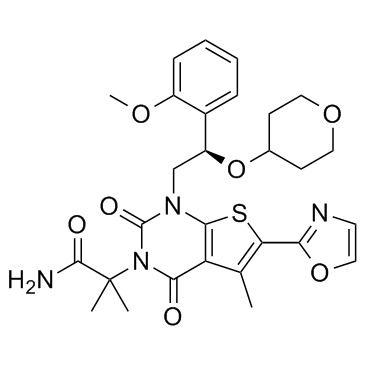
ND-646 is an orally bioavailable and steric inhibitor of acetyl-CoA carboxylase (ACC) with IC50s of 3.5 nM and 4.1 nM for recombinant hACC1 and hACC2, respectively.
|
| Related Catalog |
|
| Target |
IC50: 3.5 nM (hACC1), 4.1 nM (hACC2)[1]
|
| In Vitro |
ND-646 inhibits both ACC1 and ACC2 and therefore precludes the ability of ACC2 to compensate for ACC1 inhibition. ND-646 inhibits dimerization of recombinant human ACC2 BC domain (hACC2-BC) under native conditions; hACC2-BC migrates as a dimer in its absence and a monomer in its presence. In cell free systems, ND-646 inhibits enzymatic activity of recombinant human ACC1 (hACC1) with an IC50 of 3.5 nM and recombinant human ACC2 (hACC2) with an IC50 of 4.1 nM[1].
|
| In Vivo |
To explore the impact of chronic ND-646 treatment on NSCLC tumor growth and to determine the efficacy of twice-daily dosing, athymic nude mice bearing established A549 subcutaneous tumors are treated orally with either vehicle twice daily (BID), 25 mg/kg ND-646 once daily (QD), 25 mg/kg ND-646 BID or 50 mg/kg ND-646 QD for 31 days. ND-646 at 25 mg/kg QD is ineffective at inhibiting tumor growth. However, ND-646 administered at 25 mg/kg BID or 50 mg/kg QD significantly inhibits subcutaneous A549 tumor growth. ND-646 is well tolerated throughout the treatment period, with no significant weight loss occurring after chronic ND-646 dosing, suggesting that the maximum tolerated dose (MTD) has not been reached. Mice are sacrificed at 1 hr post final dose and tissues are either prepared for immunohistochemistry (IHC) or immunoblot analysis. Tumors treated with all doses of ND-646 have lost detection of P-ACC at 1 hr, demonstrating effective tumor penetration and acute ACC inhibition by ND-646. Notably, only at the doses of ND-646 that lead to significant tumor growth inhibition (25 mg/kg BID and 50 mg/kg QD) is significant elevation of P-EIF2αS51 expression observed in tumor lysates[1].
|
| Cell Assay |
A549, H460, H157 and H1355 cells are tested for Mycoplasma and are deemed negative. Cells are grown in DMEM plus 10% fetal bovine serum. ACC1-KO clones are maintained in 10% FBS+addition of 200 μM palmitate every 3 days. For proliferation assays via cell counts, cells are plated into 24 well plates in triplicate at 2E4 cells/well and the following day treated with either DMSO vehicle or ND-608 or ND-646. Cell counts are recorded at days 1, 3, 5 and 7-post treatment. For delipidated media, cells are first seeded in media containing regular 10% FBS and the following day are switched into media containing 20% delipidated FBS upon treatment. Viability assays are performed using either a WST-1 viability assay or Cyquant in 96 well culture plates[1].
|
| Animal Admin |
Mice[1] Subcutaneous tumor studies: For ACC1-KO studies, 8 week old athymic female nude mice are injected subcutaneously into the hind flanks with 2E6 cells of either WT or ACC1-KO luciferase expressing clones in a volume of 200 μL and imaged by BLI 20 mins after injection. Mice undergo BLI weekly for a period of 42 days (A549) and 49 days (H157), then mice are sacrificed and tumors are dissected and weighed. For ND-646 subcutaneous studies, female athymic nude mice are injected subcutaneously into the hind flanks with 5E6 A549 cells. Tumor growth is measured daily using digital calipers and volumes are calculated. Tumor prevention studies begin at 11 days post tumor cell injection. Mice are randomized into their treatment groups based on similar average tumor volumes. The average tumor volume at the initiation of treatment is 40 mm3. Mice are dosed orally (Per OS) once or twice a day with either a vehicle solution or ND-646 at either 25 mg/kg or 50 mg/kg (0.9% NaCl, 1% Tween 80, 0.5% methylcellulose). At the end of the study, mice are euthanized 1hr post final dose.
|
| References |
[1]. Svensson RU, et al. Inhibition of acetyl-CoA carboxylase suppresses fatty acid synthesis and tumor growth of non-small-cell lung cancer in preclinical models. Nat Med. 2016 Oct;22(10):1108-1119.
|
