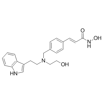
404951-53-7
| Name | Dacinostat |
|---|---|
| Synonyms |
2-Propenamide, N-hydroxy-3-[4-[[(2-hydroxyethyl)[2-(1H-indol-3-yl)ethyl]amino]methyl]phenyl]-, (2E)-
Dacinostat LAQ-824 (2E)-N-Hydroxy-3-[4-({(2-hydroxyethyl)[2-(1H-indol-3-yl)ethyl]amino}methyl)phenyl]acrylamide (E)-N-hydroxy-3-[4-[[2-hydroxyethyl-[2-(1H-indol-3-yl)ethyl]amino]methyl]phenyl]prop-2-enamide (2E)-N-hydroxy-3-[4-({(2-hydroxyethyl)[2-(1H-indol-3-yl)ethyl]amino}methyl)phenyl]prop-2-enamide NVP-LAQ824 UNII-V10P524501 S1095_Selleck |
| Description | Dacinostat is a potent HDAC inhibitor, with an IC50 of 32 nM; Dacinostat also inhibits HDAC1 with an IC50 of 9 nM, and used in cancer research. |
|---|---|
| Related Catalog | |
| Target |
HDAC1:9 nM (IC50) HDAC:32 nM (IC50) |
| In Vitro | Dacinostat (NVP-LAQ824) activates p21 promoter, with AC50 of 0.30 μM. NVP-LAQ824 inhibits tumor cell (H1299, HCT116) growth, with IC50s of 150 and 10 nM, respectively. NVP-LAQ824 also shows inhibitory activities against two prostate cancer cell lines (DU145 and PC3) and a breast cancer line (MDA435), with IC50s of 18, 23, 39 nM, respectively. Continuous exposure of NVP-LAQ824 for 72 h produces LD90s of 0.09 M in HCT116 cells and 0.47 M in A549 cells. NVP-LAQ824 treatment of NDHF cells causes the expected G1-S growth arrest in addition to a significant reduction of cells in S-phase and accumulation of cells at the G2-M checkpoint. NVP-LAQ824 induces apoptotic death in human tumor cells. NPV-LAQ824 increases acetylation of histones H3 and H4[1]. Dacinostat inhibits HDAC1 with an IC50 of 9 nM[2]. Dacinostat (10 and 20 nM) suppresses proliferation of AML fusion protein-expressing 32D cells. Dacinostat impairs short-term engraftment potential of leukemic stem cells. Dacinostat exhausts in vitro self-renewal potential of murine AML1/ETO- and PLZF/RARα-positive HSC[3]. |
| In Vivo | NVP-LAQ824 produces a dose-dependent inhibition of tumor growth, and at 100 mg/kg, its antitumor effect is similar to that of 5-Fluorouracil[1]. |
| Kinase Assay | The HDAC enzymatic assay measures compound activity in inhibiting purified HDAC isoforms. HDACs 1, 3, and 6 are immunopurified from 293 cells stably expressing the FLAG-tagged HDAC isoform, whereas HDACs 2, 4, 5, 7, 8, 9, 10, and 11 are purified from the baculovirus expression system. HDAC activity is measured in a fluorescent assay in which deacetylation of the substrate, bis-Boc-(Ac)Lys-rhodamine 110, generates a fluorophore that can be detected on a fluorometric plate reader[2]. |
| Cell Assay | Cells are plated at 5000−10000 cells per well in 96-well plates and treated with eight serial compound dilutions. Cell viability following 72 h of compound treatment is measured using the CellTiter-Glo or MTS assay. XLfit 4 is used for plotting of the growth curves and calculation of IC50 values[2]. |
| Animal Admin | The studies are performed on-site, using outbred athymic (nu/nu) female mice. Mice are anesthetized with Metofane, and a cell suspension (100 μL) containing 1×10⁶ HCT116 cells is injected s.c. into the right axillary (lateral) region of each animal. Tumors are allowed to reach the volume of approximately 100-400 mm3. At this point, mice bearing tumors with acceptable morphology (non-necrotic) and of similar size range are selected and distributed into groups of six for the studies. NVP-LAQ824 is dissolved in DMSO to create a stock solution, which is further diluted just before dosing with D5W to a final DMSO concentration of 10%. Tumor-bearing mice are treated with the compound by i.v. injection into the tail vein. NVP-LAQ824 is dosed once daily, 5 days/week, for a total of 15 doses. 5-Fluorouracil is administered at 100 mg/kg in 0.9% saline 1 day/week for a total of three doses. The control groups are treated with the vehicle. Tumors are collected from the animals at the indicated time points[1]. |
| References |
| Density | 1.3±0.1 g/cm3 |
|---|---|
| Molecular Formula | C22H25N3O3 |
| Molecular Weight | 379.452 |
| Exact Mass | 379.189606 |
| PSA | 88.59000 |
| LogP | 3.41 |
| Index of Refraction | 1.693 |
| Storage condition | -20℃ |
