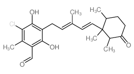
| In Vitro |
Ascochlorin (Ilicicolin D) (10-50 μM; 24-72 hours) inhibits the viability of HepG2, HCCLM3 and Huh7 cells in a time and dose dependent manner[3]. Ascochlorin (50 μM; 48 hours) induces apoptosis in HCC cells[3]. Ascochlorin (1-50 μM) significantly suppresses the production of nitric oxide (NO) and prostaglandin E2 (PGE2) and decreases the gene expression of inducible NO synthase (iNOS) and cyclooxygenase-2 (COX-2) in a dose-dependent manner. Ascochlorin inhibits the mRNA expression and the protein secretion of interleukin (IL)-1β and IL-6 but not tumor necrosis factor (TNF)-α in LPS-stimulated RAW 264.7 macrophage cells. Ascochlorin suppresses nuclear translocation and DNA binding affinity of nuclear factor-κB (NF-κB). Ascochlorin down-regulates phospho-extracellular signal-regulated kinase 1/2 (p-ERK1/2) and p-p38[2]. Cell Viability Assay[3] Cell Line: HepG2, HCCLM3, Huh7 cells Concentration: 10, 25, 50 μM Incubation Time: 24, 48, 72 hours Result: Inhibit the viability of three different HCC cell lines tested (HepG2, HCCLM3 and Huh7) in a time and dose dependent manner. Western Blot Analysis[3] Cell Line: HepG2 cells Concentration: 50 μM Incubation Time: 48 hours Result: Inhibited expression of the cell cycle regulator protein cyclin D1, the anti-apoptotic proteins Bcl-2, Mcl-1, survivin and XIAP, and the invasive gene product MMP-9.
|
| In Vivo |
Ascochlorin (Ilicicolin D) (2.5-5 mg/kg; i.p.; day 0, 1, 2, 3, 13, 15, 17, 20, 22, 24, 27, 29 and 31) inhibits tumor growth in an orthotopic HCC mouse model[1]. Animal Model: Eight week-old athymic balb/c nude female mice (HCCLM3-Luc2 tumors)[3] Dosage: 2.5 mg/kg, 5 mg/kg Administration: I.p.; day 0, 1, 2, 3, 13, 15, 17, 20, 22, 24, 27, 29 and 31 Result: Induced significant inhibition of tumor growth.
|
