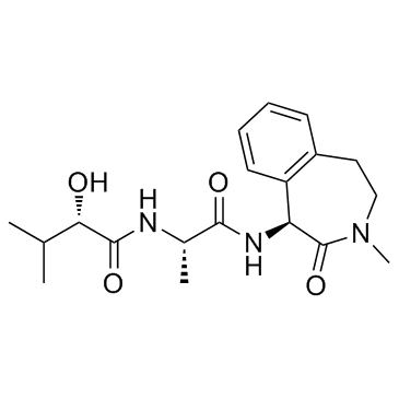
425386-60-3
| Name | Semagacestat |
|---|---|
| Synonyms |
LY450139
(2S)-2-Hydroxy-3-methyl-N-[(2S)-1-{[(1S)-3-methyl-2-oxo-2,3,4,5-tetrahydro-1H-3-benzazepin-1-yl]amino}-1-oxopropan-2-yl]butanamide (2S)-2-Hydroxy-3-methyl-N-[(2S)-1-{[(1S)-3-methyl-2-oxo-2,3,4,5-tetrahydro-1H-3-benzazepin-1-yl]amino}-1-oxopropan-2-yl]butanamid Semagacestat (2S)-2-Hydroxy-3-methyl-N-[(2S)-1-{[(1S)-3-methyl-2-oxo-2,3,4,5-tetrahydro-1H-3-benzazepin-1-yl]amino}-1-oxo-2-propanyl]butanamide Butanamide, 2-hydroxy-3-methyl-N-[(1S)-1-methyl-2-oxo-2-[[(1S)-2,3,4,5-tetrahydro-3-methyl-2-oxo-1H-3-benzazepin-1-yl]amino]ethyl]-, (2S)- |
| Description | Semagacestat is a γ-secretase inhibitor, inhibits β-amyloid (Aβ42), Aβ38 and Aβ40 with IC50 of 10.9, 12 and 12.1 nM, respectively; also inhibits Notch signaling with IC50 of 14.1 nM. |
|---|---|
| Related Catalog | |
| Target |
IC50: 10.9 nM (Aβ42), 12 nM (Aβ38), 12.1 nM (Aβ40), 14.1 nM (Notch)[1] |
| In Vitro | Semagacestat (LY450139) reduces the secretion of Aβ42, Aβ40, and Aβ38 in 96-well-cultured media and increases β-CTF in cell lysates as expected, although this increase is unexpectedly attenuated at high concentrations[1]. In cortical neurons (CTX), Semagacestat (LY450139) causes a concentration-dependent decrease in Aβ40 secreted into the medium with IC50 value 111 nM for Semagacestat. Semagacestat causes a concentration-dependent decrease in Aβ40 and Aβ42 secreted into the medium with an IC50 value of 126 and 130 nM, respectively[2]. |
| In Vivo | Semagacestat (LY450139) is found to decrease both Aβ42 and Aβ40 at 10 mg/kg (22-23% reduction;p<0.01) and increase β-CTF at 0.3-10 mg/kg in a dose-dependent manner (15-162% elevation; p<0.01 at 1-10 mg/kg)[1]. The γ-secretase inhibitor, Semagacestat (LY450139), a highly potent low molecular weight compound, significantly reduces β-amyloid (Aβ) levels in cell cultures permanently over-expressing APP and in both wildtype and transgenic APP-expressing mice. Three hours following p.o. dosing of 30 mg/kg Semagacestat levels of Aβ40 are reduced by 43% (unpaired t-test, p=0.002) in the brains of wildtype C57BL/6 mice compare with vehicle treated controls. Subcutaneous administration of Semagacestat (30 mg/kg) transiently decreases the amounts of Aβ40 in the dialysate with a maximum reduction in Aβ40 levels of 80% at 3 h post-dosing (p<0.001)[2]. |
| Kinase Assay | H4 human glioma cells stably overexpressing human wild-type APP695 are maintained in DMEM supplemented with 10% fetal bovine serum and penicillin/streptomycin. Cells are cultured in 96- or 6-well plates overnight, and then treated with each drug (e.g., Semagacestat) at various concentrations for 24 h. Levels of Aβ1-42, Aβ1-40, and Aβ1-38 in the media are measured using separate ELISA kits. To quantify β-CTF, cells are lysed with RIPA buffer (25 mM Tris, 150 mM NaCl, 1% NP-40, 1% sodium deoxycholate, 0.1% SDS; pH 7.6) containing Complete protease inhibitor mixture and applied to a human β-CTF ELISA kit at 1:20 dilution. Aliquots of the cell lysate are also used for CellTiter-Glo Luminescent Cell Viability Assay. The cell lysate from the six-well plate is subjected to Western blot analysis[1]. |
| Cell Assay | Murine cortical neurons (CTX) are isolated from day 14 to 16 foetal C57BL/6 mice. Briefly, dissociated neurons are plated on 100 μg/mL poly-L-lysine coated dishes at a density of 0.25×10⁶ cells/cm2 (800000 cells/mL; 100 μL/well, 96-well plate) and cultured in Neurobasal medium supplemented with 2% B-27 supplement without antioxidants, 0.5 mM L-glutamine and 100 U/mL penicillin and 0.1 mg/mL streptomycin. Neurons are fed every third day by replacing half of the medium. The proportion of glia cells in the cultures is less than 10%, as assessed by an antibody against glia-fibrillary acidic protein. CTX are used at 6 days in vitro (DIV) after complete medium change and incubated with secretase inhibitors (e.g., Semagacestat) for 24 h. Neurons and cell medium are used at DIV 7. For detection of cell viability, the percentage of viable cells is quantified by their capacity to reduce MTT following incubation with 0.5 mg/mL MTT for 60 min. Viability is routinely measured after all in vitro pharmacological experiments[2]. |
| Animal Admin | Mice[1] Female Tg2576 mice expressing human APP695 with the Swedish mutation (K670N/M671L) are used. Male transgenic mice are procured and crossbred with female B6SJLF1/J mice. To identify drug effects on cognitive function, four different experiments are conducted. The objective of Experiment 1 is to elucidate acute and subchronic drug effects on cognitive deficits in Tg2576 mice. Each drug (Semagacestat, BMS-708163, and GSM-2) is orally administered to 5.5-month-old Tg2576 mice for 8 d. Y-maze tests are conducted to evaluate spatial working memory 3 h after administration on days 1 and 8. Vehicle-treated Tg2576 mice demonstrates significantly lower spontaneous alternation rates than WT mice in the Y-maze test, suggesting deficits in spatial working memory. On day 1, 1 mg/kg Semagacestat, 1 mg/kg BMS-708163, and 0.1-0.3 mg/kg GSM-2 significantly ameliorates these cognitive deficits (acute effects). On day 8, however, the GSI effects disappear, whereas GSM-2 retained its significant effects (subchronic effects). Mice are killed immediately after the Y-maze test on day 8, when hippocampal levels of Aβ42, Aβ40, and β-CTF are determined by ELISA. |
| References |
| Density | 1.2±0.1 g/cm3 |
|---|---|
| Boiling Point | 681.9±55.0 °C at 760 mmHg |
| Melting Point | 208-212°C |
| Molecular Formula | C19H27N3O4 |
| Molecular Weight | 361.435 |
| Flash Point | 366.2±31.5 °C |
| Exact Mass | 361.200165 |
| PSA | 98.74000 |
| LogP | 2.23 |
| Vapour Pressure | 0.0±2.2 mmHg at 25°C |
| Index of Refraction | 1.576 |
| Storage condition | 2-8°C |
