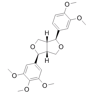
31008-18-1
| Name | magnolin |
|---|---|
| Synonyms |
5-methoxyeudesmin
Maglin (1S,3aR,4S,6aR)-1-(3,4-Dimethoxyphenyl)-4-(3,4,5-trimethoxyphenyl)tetrahydro-1H,3H-furo[3,4-c]furan (7S,7’S,8R,8’R)-magnolin 1H,3H-Furo[3,4-c]furan, 1-(3,4-dimethoxyphenyl)tetrahydro-4-(3,4,5-trimethoxyphenyl)-, (1S,3aR,4S,6aR)- medioresinol dimethyl ether |
| Description | Magnolin, a major component of Magnolia flos (Shin-Yi), inhibits the Ras/ERKs/RSK2 signaling axis by targeting the active pocket of ERK1 and ERK2 with IC50s of 87 nM and 16.5 nM, respectively. |
|---|---|
| Related Catalog | |
| Target |
ERK2:16.5 nM (IC50) ERK1:87 nM (IC50) |
| In Vitro | Magnolin is a natural compound abundantly found in Magnolia flos, which has been traditionally used in oriental medicine to treat headaches, nasal congestion and anti-inflammatory reactions. Magnolin targets the active pockets of ERK1 and ERK2, which are important signaling molecules in cancer cell metastasis. Magnolin inhibits NF-κB transactivation activity by suppressing the ERKs/RSK2 signaling pathway. Magnolin inhibits the production of tumor necrosis factor-α (TNF-α) and prostaglandin E2 (PGE2) by inhibiting extracellular signal-regulated kinases (ERKs), which are key signaling molecules in the regulation of cell proliferation, transformation and cancer cell metastasis. JB6 Cl41 cell migration enhanced by EGF treatment is dramatically suppressed by Magnolin treatment in a dose-dependent manner. Magnolin inhibits ERK1/2/RSK2 signaling-mediated IκBα phosphorylation at Ser32, resulting in the inhibition of NF-κB activation and cell migration[1]. |
| Cell Assay | JB6 Cl41 (7×10⁴), A549 (7×10⁴) and NCI-H1975 (7×10⁴) cells, and RSK2+/+ (7×10⁴) and RSK2-/- (7×10⁴) MEFs are seeded into culture-inserts and cultured overnight. The cells are treated with mitomycin-C (10 μg/mL) for 2 h, and the culture-inserts are removed to offer a cell-free gap. The cells are treated with the indicated doses of Magnolin (15, 30, and 60 μM) either in the presence or absence of EGF for 12 or 24 h, and cell migration is observed under a light microscope. The migrated area is measured using the Image J computer software program. To measure the Magnolin effect on cancer cell invasion, a matrigel-coated invasion chamber is used. Briefly, A549 or NCI-H1975 (2.5×10⁴) cells are seeded into an insert chamber with FBS-free media supplemented with the indicated doses of Magnolin(15, 30, and 60 μM), and cultured in 24-well plates supplemented with complete media for the appropriate time period. The cells are fixed with 4 % formaldehyde, permeabilized with methanol and stained with crystal violet. The stained cells are observed under a light microscope and those that have migrated are counted[1]. |
| References |
| Density | 1.2±0.1 g/cm3 |
|---|---|
| Boiling Point | 536.9±50.0 °C at 760 mmHg |
| Molecular Formula | C23H28O7 |
| Molecular Weight | 416.464 |
| Flash Point | 215.6±30.0 °C |
| Exact Mass | 416.183502 |
| PSA | 64.61000 |
| LogP | 2.78 |
| Vapour Pressure | 0.0±1.4 mmHg at 25°C |
| Index of Refraction | 1.542 |

