| Description |
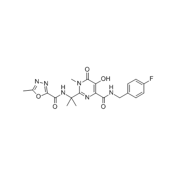
Raltegravir is a potent integrase (IN) inhibitor, used to treat HIV infection.
|
| Related Catalog |
|
| In Vitro |
PFV IN carrying the S217H substitution is 10-fold less susceptible to Raltegravir with IC50 of 900 nM. PFV IN displays 10% of WT activity and is inhibited by Raltegravir with an IC50 of 200 nM, indicating a appr twofold decrease in susceptibility to the IN strand transfer inhibitor (INSTI) compared with WT IN. S217Q PFV IN is as sensitive to Raltegravir as the WT enzyme[1]. Raltegravir is metabolized by glucuronidation, not hepatically. Raltegravir has potent in vitro activity against HIV-1, with a 95% inhibitory concentration of 31±20 nM, in human T lymphoid cell cultures. Raltegravir is also active against HIV-2 when Raltegravir is tested in CEMx174 cells, with an IC95of 6 nM. Raltegravir metabolism occurs primarily through glucuronidation. Drugs that are strong inducers of the glucuronidation enzyme, UGT1A1, significantly reduce Raltegravir concentrations and should not be used. Raltegravir exhibits weak inhibitory effects on hepatic cytochrome P450 activity. Raltegravir does not induce CYP3A4 RNA expression or CYP3A4-dependent testosterone 6-β-hydroxylase activity[2]. Raltegravir cellular permeativity is reduced in the presence of magnesium and calcium[3]. Raltegravir and related HIV-1 integrase (IN) strand transfer inhibitors (INSTIs efficiently block viral replication[4]. In acutely infected human lymphoid CD4+ T-cell lines MT-4 and CEMx174, SIVmac251 replication is efficiently inhibited by Raltegravir, which shows an EC90 in the low nanomolar range[5].
|
| In Vivo |
Raltegravir induces viro-immunological improvement of nonhuman primates with progressing SIVmac251 infection. One non-human primate shows an undetectable viral load following Raltegravir monotherapy[5].
|
| Kinase Assay |
For quantitative strand transfer assays, donor DNA substrate is formed by annealing HPLC grade oligonucleotides 5'-GACTCACTATAGGGCACGCGTCAAAATTCCATGACA and 5'-ATTGTCATG GAATTTTGACGCGTGCCCTATAGTGAGTC. Reactions (40 μL) contains 0.75 μM PFV IN, 0.75 μM donor DNA, 4 nM (300 ng) supercoiled pGEM9-Zf(−) target DNA, 125 mM NaCl, 5 mM MgSO4, 4 μM ZnCl2, 10 mM DTT, 0.8% (vol/vol) DMSO, and 25 mM BisTris propane-HCl, pH 7.45. Raltegravir is added at indicated concentrations. Reactions are initiated by addition of 2 μL PFV IN diluted in 150 mM NaCl, 2 mM DTT, and 10 mM Tris-HCl, pH 7.4, and stopped after 1 hour at 37°C by addition of 25 mM EDTA and 0.5% (wt/vol) SDS. Reaction products, deproteinized by digestion with 20 μg proteinase K for 30 minutes at 37°C followed by ethanol precipitation, are separated in 1.5% agarose gels and visualized by staining with ethidium bromide. Integration products are quantified by quantitative real-time PCR, using Platinum SYBR Green qPCR SuperMix and three primers: 5'-CTACTTACTCTAGCTTCCCGGCAAC, 5'-TTCGCCAGTTAATAGTTTGCGCAAC, and 5'-GACTCACTATAGGGCACGCGT. PCR reactions (20 μL) contained 0.5 μM of each primer and 1 μL diluted integration reaction product. Following a 5-min denaturation step at 95°C, 35 cycles are carried out in a CFX96 PCR instrument, using 10 seconds denaturation at 95°C, 30 seconds annealing at 56°C and 1 minutes extension at 68°C. Standard curves are generated using serial dilutions of WT PFV IN reaction in the absence of INSTI.
|
| Cell Assay |
Human MT-4 cells are infected for 2 hours with the SIVmac251, HIV-1 (IIIB) and HIV-2 (CDC 77618) stocks at a multiplicity of infection of, approximately, 0.1. Cells are then washed three times in phosphate buffered saline, and suspended at 5 × 10⁵/mL in fresh culture medium (to primary cells 50 units/mL of IL-2 are added) in 96-well plates, in the presence or absence of a range of triplicate raltegravir concentrations (0.0001 μM-1 μM). Untreated infected and mock-infected controls are prepared too, in order to allow comparison of the data derived from the different treatments. Viral cytopathogeniciy in MT-4 cells is quantitated by the methyl tetrazolium (MTT) method (MT-4/MTT assay) when extensive cell death in control virus-infected cell cultures is detectable microscopically as lack of capacity to re-cluster. The capability of MT-4 cells to form clusters after infection. Briefly, clusters are disrupted by pipetting; and, after 2 hours of incubation at 37°C, the formation of new clusters is assessed by light microscopy (100× magnification). Cell culture supernatants are collected for HIV-1 p24 and HIV-2/SIVmac251 p27 core antigen measurement by ELISA. In CEMx174-infected cell cultures, which show a propensity to form syncytia induced by the virus envelope glycoproteins, syncytia are counted, in blinded fashion, by light microscopy for each well at 5 days following infection.
|
| References |
[1]. Hare, S., et al., Molecular mechanisms of retroviral integrase inhibition and the evolution of viral resistance. Proc Natl Acad Sci U S A, 2010. 107(46): p. 20057-62. [2]. Hicks C, et al. Raltegravir: the first HIV type 1 integrase inhibitor. Clin Infect Dis. 2009 Apr 1;48(7):931-9 [3]. Moss DM, et al. Divalent metals and pH alter raltegravir disposition in vitro. Antimicrob Agents Chemother. 2012 Jun;56(6):3020-6 [4]. Hare S, et al. Structural and functional analyses of the second-generation integrase strand transfer inhibitor dolutegravir (S/GSK1349572). Mol Pharmacol. 2011 Oct;80(4):565-72. [5]. Lewis, M.G., et al. Response of a simian immunodeficiency virus (SIVmac251) to raltegravir: a basis for a new treatment for simian AIDS and an animal model for studying lentiviral persistence during antiretroviral therapy. Retrovirology, 2010. 7: p. 21. [6]. Xu P, et al. Combined Medication of Antiretroviral Drugs Tenofovir Disoproxil Fumarate, Emtricitabine, and Raltegravir Reduces Neural Progenitor Cell Proliferation In Vivo and In Vitro. J Neuroimmune Pharmacol. 2017 Dec;12(4):682-692.
|
