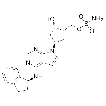
905579-51-3
| Name | [(1S,2S,4R)-4-{4-[(1S)-2,3-Dihydro-1H-inden-1-ylamino]-7H-pyrrolo [2,3-d]pyrimidin-7-yl}-2-hydroxycyclopentyl]methyl sulfamate |
|---|---|
| Synonyms |
MLN 4924
Pevonedistat MLN4924 [(1S,2S,4R)-4-{4-[(1S)-2,3-Dihydro-1H-inden-1-ylamino]-7H-pyrrolo[2,3-d]pyrimidin-7-yl}-2-hydroxycyclopentyl]methyl sulfamate [(1S,2S,4R)-4-(4-{[(1S)-2,3-dihydro-1H-inden-1-yl]amino}-7H-pyrrolo[2,3-d]pyrimidin-7-yl)-2-hydroxycyclopentyl]methyl sulfamate MLN-4924 |
| Description | MLN4924 is a potent and selective NEDD8-activating enzyme (NAE) inhibitor with an IC50 of 4.7 nM. |
|---|---|
| Related Catalog | |
| Target |
IC50: 4.7 nM (NAE)[1] |
| In Vitro | MLN4924 is a potent inhibitor of NAE, and is selective relative to the closely related enzymes UAE, SAE, UBA6 and ATG7 (IC50=1.5, 8.2, 1.8 and >10 μM, respectively) when evaluated in purified enzyme assays that monitor the formation of E2-UBL thioester reaction products. MLN4924 selectively inhibits NAE activity compared to the closely related ubiquitin-activating enzyme (UAE, also known as UBA1) and SUMO-activating enzyme (SAE; a heterodimer of SAE1 and UBA2 subunits), in purified enzyme and cellular assays. MLN4924 exhibits potent cytotoxic activity against a variety of human tumour-derived cell lines[1]. |
| In Vivo | MLN4924 (sc, 10 mg/kg, 30 mg/kg, or 60 mg/kg) inhibits the NEDD8 pathway resulting in DNA damage in Mice bearing HCT-116 xenografts[1].Pevonedistat (sc, 120 mg/kg) and TNF-α (10 μg/kg) synergistically cause liver damage in SD rats[2]. |
| Cell Assay | HCT-116 cells grown in 6-well cell-culture dishes are treated with 0.1% DMSO (control) or 0.3 μM MLN4924 for 24 h. Whole cell extracts are prepared and analysed by immunoblotting. For analysis of the E2-UBL thioester levels, lysates are fractionated by non-reducing SDS-PAGE and immunoblotted with polyclonal antibodies to Ubc12, Ubc9 and Ubc10. For analysis of other proteins, lysates are fractionated by reducing SDS-PAGE and probed with primary antibodies as follows: mouse monoclonal antibodies to CDT1, p27, geminin, ubiquitin, securin/PTTG and p53 or rabbit polyclonal antibodies to NRF2, Cyclin B1 and GADD34[1]. |
| Animal Admin | Mice[1] Mice bearing HCT-116 tumours of 300-500 mm3 are administered a single MLN4924 dose (of 10, 30 or 60 mg/kg), and tumors are excised at various time-points over the subsequent 24 h period. The relative levels of NEDD8-cullin and NRF2 are estimated by quantitative immunoblot analysis using Alexa680-labelled anti-IgG as the secondary antibody. The statistical difference between the groups for NEDD8-cullin inhibition is determined using the Kruskal-Wallis test. For the analysis of CDT1 and phosphorylated CHK1 (Ser317) levels in tumour sections, formalin-fixed, paraffin-embedded tumour sections are stained with the relevant antibodies, amplified with HRP-labelled secondary antibodies and detected with the ChromoMap DAB Kit. Slides are counterstained with haematoxylin. Images are captured using an Eclipse E800 microscope and Retiga EXi colour digital camera and processed using Metamorph software. CDT1 and phosphorylated CHK1 levels are expressed as a function of the DAB signal area. Rats[2] Ten-week-old male Sprague-Dawley rats are used. Across two studies, a total of eight animals in each group are dosed with vehicle, TNF-α, MLN4924, or MLN4924+TNF-α. Animals are first intravenously administered either vehicle (1×PBS) or 10 μg/kg TNF-α. One hour later, they are subcutaneously administered vehicle (20% sulfobutyl ether beta-cyclodextrin in 50 mM citrate buffer, pH 3.3) or 120 mg/kg MLN4924. Scheduled euthanasia occurred 24 h postdose. Unscheduled euthanasia is performed when animals exhibited moribund conditions. Serum is collected at necropsy and analyzed by Idexx Laboratories for serum chemistry markers of liver damage. Additionally, the livers from five animals in each group are removed, separated into two sections and either frozen at -80°C for subsequent protein analysis or fixed in 10% neutral buffered formalin, embedded in paraffin, sectioned at 4-6 μm, mounted on glass slides, stained with hematoxylin and eosin, and analyzed with an Olympus BX51 light microscope for histopathology assessment. Microscopic findings are recorded in concordance with the standardized nomenclature for classifying lesions within the livers of rats. |
| References |
| Density | 1.6±0.1 g/cm3 |
|---|---|
| Boiling Point | 721.0±70.0 °C at 760 mmHg |
| Melting Point | 161-163°C |
| Molecular Formula | C21H25N5O4S |
| Molecular Weight | 443.519 |
| Flash Point | 389.9±35.7 °C |
| Exact Mass | 443.162720 |
| PSA | 140.74000 |
| LogP | 2.16 |
| Vapour Pressure | 0.0±2.4 mmHg at 25°C |
| Index of Refraction | 1.769 |
| Storage condition | -20?C Freezer |
