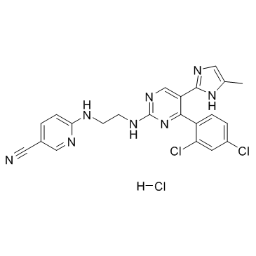
CHIR-99021 HCl
Modify Date: 2024-01-11 17:45:11

CHIR-99021 HCl structure
|
Common Name | CHIR-99021 HCl | ||
|---|---|---|---|---|
| CAS Number | 1797989-42-4 | Molecular Weight | 501.799 | |
| Density | N/A | Boiling Point | N/A | |
| Molecular Formula | C22H19Cl3N8 | Melting Point | N/A | |
| MSDS | N/A | Flash Point | N/A | |
Use of CHIR-99021 HClCHIR-99021 monohydrochloride is a GSK-3α/β inhibitor with IC50 of 10 nM/6.7 nM; > 500-fold selectivity for GSK-3 versus its closest homologs CDC2 and ERK2, as well as other protein kinases. |
| Name | CHIR-99021 monohydrochloride |
|---|---|
| Synonym | More Synonyms |
| Description | CHIR-99021 monohydrochloride is a GSK-3α/β inhibitor with IC50 of 10 nM/6.7 nM; > 500-fold selectivity for GSK-3 versus its closest homologs CDC2 and ERK2, as well as other protein kinases. |
|---|---|
| Related Catalog | |
| Target |
GSK-3β:6.7 nM (IC50) GSK-3α:10 nM (IC50) cdc2:8800 nM (IC50) |
| In Vitro | CHIR 99021inhibits human GSK-3β with Ki values of 9.8 nM[1]. CHIR 99021 is a small organic molecule that inhibits GSK3α and GSK3β by competing for their ATP-binding sites.In vitro kinase assays reveal that CHIR 99021 specifically inhibits GSK3β (IC50=~5 nM) and GSK3α (IC50=~10 nM), with little effect on other kinases[2]. In the presence of CHIR-99021 the viability of the ES-D3 cells is reduced by 24.7% at 2.5 μM, 56.3% at 5 μM, 61.9% at 7.5 μM and 69.2% at 10 μM CHIR-99021 with an IC50 of 4.9 μM[3]. |
| In Vivo | In ZDF rats, a single oral dose of CHIR 99021 (16 mg/kg or 48 mg/kg) rapidly lowers plasma glucose, with a maximal reduction of nearly 150 mg/dl 3-4 h after administration[1]. CHIR99021 (2 mg/kg) given once, 4 h before irradiation, significantly improves survival after 14.5 Gy abdominal irradiation (ABI). CHIR99021 treatment significantly blocks crypt apoptosis and accumulation of p-H2AX+ cells, and improves crypt regeneration and villus height. CHIR99021 treatment increases Lgr5+ cell survival by blocking apoptosis, and effectively prevents the reduction of Olfm4, Lgr5 and CD44 as early as 4 h[4]. |
| Kinase Assay | Kinases are purified from SF9 cells through use of their His or Glu tag. Glu-tagged proteins are purified, and His-tagged proteins are purified. Kinase assays are performed in 96-well plates with appropriate peptide substrates in a 300-μL reaction buffer (variations on 50 mM Tris-HCl, pH 7.5, 10 mM MgCl2, 1 mM EGTA, 1 mMdithiothreitol, 25 mMβ-glycerophosphate, 1 mM NaF, and 0.01% bovine serum albumin). Peptides has Km values from 1 to 100 μM. CHIR 99021 or CHIR GSKIA is added in 3.5 μL of Me2SO, followed by ATP to a final concentration of 1 μM. After incubation, triplicate 100-μL aliquots are transferred to Combiplate 8 plates containing 100 μL/well of 50 μM ATP and 20 mM EDTA. After 1 hour, the wells are rinsed five times with phosphate-buffered saline, filled with 200 μL of scintillation fluid, sealed, and counted in a scintillation counter 30 min later. All of the steps are at room temperature. The percentage of inhibition is calculated as 100×(inhibitor-no enzyme control)/(Me2SO control-no enzyme control)[2]. |
| Cell Assay | The viability of the mouse ES cells is determined after exposure to different concentrations of GSK3 inhibitors for three days using the MTT assay. The decrease of MTT activity is a reliable metabolism-based test for quantifying cell viability; this decrease correlates with the loss of cell viability. 2,000 cells are seeded overnight on gelatine-coated 96-well plates in LIF-containing ES cell medium. On the next day the medium is changed to medium devoid of LIF and with reduced serum and supplemented with 0.1-1 μM BIO, or 1-10 μM SB-216763, CHIR-99021 or CHIR-98014. Basal medium without GSK3 inhibitors or DMSO is used as control. All tested conditions are analyzed in triplicates[3]. |
| Animal Admin | Rats[1] Primary hepatocytes from male Sprague Dawley rats that weighed <140 g are prepared and used 1-3 h after isolation. Aliquots of 1×10⁶cells in 1 mL of DMEM/F12 medium plus 0.2% BSA and CHIR 99021(orally at 16 or 48 mg/kg) or controls are incubated in 12-well plates on a low-speed shaker for 30 min at 37°C in a CO2-enriched atmosphere, collected by centrifugation and lysed by freeze/thaw in buffer A plus 0.01% NP40; the GS assay is again performed. Mice[4] Mice 6-10 weeks old are used. The PUMA+/+ and PUMA-/- littermates on C57BL/6 background (F10) and Lgr5-EGFP (Lgr5-EGFP-IRES-creERT2) mice are subjected to whole body irradiation (TBI), or abdominal irradiation (ABI). Mice are injected intraperitoneally (i.p.) with 2 mg/kg of CHIR99021 4 h before radiation or 1 mg/kg of SB415286 28 h and 4 h before radiation. Mice are sacrificed to collect small intestines for histology analysis and western blotting. All mice are injected i.p. with 100 mg/kg of BrdU before sacrifice. |
| References |
| Molecular Formula | C22H19Cl3N8 |
|---|---|
| Molecular Weight | 501.799 |
| Exact Mass | 500.079834 |
| Storage condition | -20℃ |
| 3-Pyridinecarbonitrile, 6-[[2-[[4-(2,4-dichlorophenyl)-5-(4-methyl-1H-imidazol-2-yl)-2-pyrimidinyl]amino]ethyl]amino]-, hydrochloride (1:1) |
| MFCD23704170 |
| 6-[(2-{[4-(2,4-Dichlorophenyl)-5-(4-methyl-1H-imidazol-2-yl)-2-pyrimidinyl]amino}ethyl)amino]nicotinonitrile hydrochloride (1:1) |
| CHIR-99021 (monohydrochloride) |