| In Vitro |
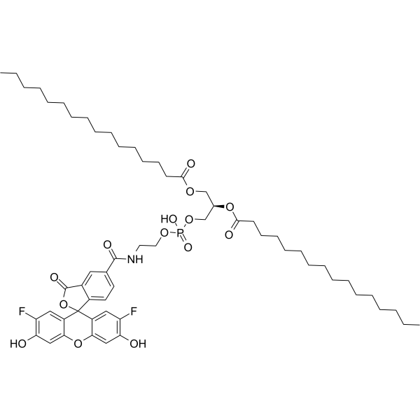
FG 488 DHPE shows a pH-dependent fluorescence emission characteristic[1]. Monitoring acidification in Bulk vesicle assa[1]: 1.Instrument: Jasco FP6500 spectrofluorometer, 37 ℃; fluorescence is excited at λex=508 nm and the emission is detected at λem=534 nm. 2.Add 100 µL proteoliposomes (cphospholipid is about 60 µM) to 680 µL ATPase buffer, containing the K+-ionophore valinomycin (5 nM) to enable a charge equilibration for transported protons. 3.Add ATP (1.2 mM) to induce proton pumping. 4.Add 1 mM NaN3 to ATP hydrolysis. 5.Add CCCP (carbonyl cyanide 3-chlorophenyl hydrazine, 0.4 µM) to deplete the proton gradient. 6.Conversion into pH-values, fluorescence intensities are normalized to the intensity obtained directly after ATP addition. FG 488 DHPE exerts function in quantification of pH changes induced by the voltage-dependent proton channel Hv1[2]. Quantification of phospholipid concentrations[2]: 1.Add Perchloric acid (70%, 200 μL) to a sample of unilamellar vesicles containing OG488-DHPE (30 μL). 2.Heat up to 220 °C for 60 min to generate inorganic phosphate. 3.Cooling down to room temperature, add 700 μL of a solution of NH4MoO4 (0.45% (w/v)) and perchloric acid (12.6% (w/v)) and 700 μL o f a 1.7% (w/v) acetic acid solution. 4.Obtain a calibration curve to know NaH2PO4 concentrations. 5.Incubated samples at 80 °C for 10 min and measure the absorption of the samples at 820 nm. 6.Calculate phospholipid concentrations of the vesicles using the calibration curve. Proton translocation assay[2]: 1.Instrument: Jasco FP6500 spectrofluorometer, 37 ℃; fluorescence is excited at λex=508 nm (3 nm band width) and the emission is detected at λem=534 nm (3 nm band width). 2.Dilute proteoliposomes composed of POPC/POPG/Chol/OG488-DHPE (54.5:25:20:0.5) in buffer A in flux buffer generating a 14-fold K+-gradient across the vesicular membrane. 3.Add valinomycin (13 nM) to cause protonation of OG488-DHPE and quench its fluorescence intensity in case of active Hv1 channels as described above. 4.Add CCCP (6 nM) to permeabilise all vesicles for protons. 5.The normalized fluorescence intensity Fnorm is plotted as a function of time. As a control for proton leakage, protein-free vesicles were used instead of proteoliposomes. For the experiments in the presence of the potential inhibitor 2GBI, dissolve the inhibitor (15 mM) in flux buffer and add (0.5-8.0 μL) to the proteoliposomes before addition of valinomycin to induce proton translocation.
|
