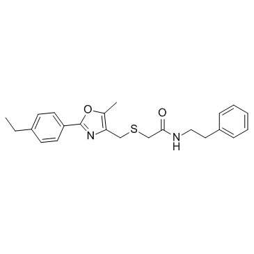
iCRT3

iCRT3 structure
|
Common Name | iCRT3 | ||
|---|---|---|---|---|
| CAS Number | 901751-47-1 | Molecular Weight | 394.53000 | |
| Density | N/A | Boiling Point | N/A | |
| Molecular Formula | C23H26N2O2S | Melting Point | N/A | |
| MSDS | Chinese USA | Flash Point | N/A | |
| Symbol |


GHS05, GHS07 |
Signal Word | Danger | |
Use of iCRT3iCRT3 is an inhibitor of both Wnt and β-catenin-responsive transcription. |
| Name | Acetamide, 2-[[[2-(4-ethylphenyl)-5-methyl-4-oxazolyl]methyl]thio]-N-(2-phenylethyl) |
|---|---|
| Synonym | More Synonyms |
| Description | iCRT3 is an inhibitor of both Wnt and β-catenin-responsive transcription. |
|---|---|
| Related Catalog | |
| Target |
Wnt |
| In Vitro | iCRT3 is an inhibitor of both Wnt and β-catenin-responsive transcription. iCRT3 significantly decreases TOP Flash activity and reduces the level of NTSR1. The anti-apoptotic effects of Neurotensin (NTS) and Wnt3a can be largely abrogated by iCRT3[1]. Cells maintained long term with iCRT3 show enhanced expression of classic pluripotency genes compare with the DMSO control, whereas expression of differentiation markers and T-cell factor (TCF) target genes is concomitantly reduced[2]. Treatment with iCRT3 at doses of 12.5, 25, 50, and 75 μM decreases TNF-α levels by 14.7%, 18.5%, 44.9% and 61.3%, respectively. With iCRT3 treatment, IκB levels are increased in a dose-dependent manner compare to the vehicle[3]. |
| In Vivo | The tumor growth rates are markedly retarded by iCRT3 treatment. Consistently, the tumor-suppressive role of iCRT3 is accompanied with a reduction in Ki67 index, a proliferation marker[1]. The IL-6 levels in the 10 mg/kg iCRT3 treatment group are 82.9% lower than those in the vehicle group. IL-1β levels are undetectable in the sham but reach 371 pg/mL in septic mice and are down by 30.2% and 53.2%, respectively, with 5 and 10 mg/kg iCRT3. With iCRT3 treatment at doses of 5 and 10 mg/kg, AST levels in these septic mice are 15.4% and 44.2% lower, respectively, than those in the vehicle-treated mice. After treatment with 10 mg/kg iCRT3, lung morphology is improved with much reduced microscopic deterioration, compare to the vehicle group. The number of apoptotic cells in the lung tissues of the iCRT3-treated mice is significantly reduced by 92.7% in comparison with the vehicle group[3]. |
| Cell Assay | Cells are seeded into 96-well plates to a density of 5×10³ cells per well and incubated in the culture medium with iCRT3 for an additional 48 h. Cell viability and cell apoptosis assays are carried out using a Cell Counting kit-8 and a Caspase-Glo 3/7 assay kit according to the manufacturer’s instructions, respectively[1]. |
| Animal Admin | NOD-SCID BALB/c mice are inoculated subcutaneously in the right back with 2×10⁶ A172 cells. The growth of the primary tumors is recorded every 4 days. iCRT3 (5 mg/kg) is diluted in PBS i.p. triweekly when tumors grow to ~200 mm3. The control mice are treated with blank PBS containing 5% (v/v) DMSO. Tumor volume is evaluated with the following formula: volume=tumor length×width2/2. The mice are sacrificed 24 days after pharmaceutical treatment. The tumors are resected and embedded in paraffin, and the Ki67 staining is analyzed by immunohistochemistry[1]. |
| References |
| Molecular Formula | C23H26N2O2S |
|---|---|
| Molecular Weight | 394.53000 |
| Exact Mass | 394.17100 |
| PSA | 80.43000 |
| LogP | 5.19540 |
| Storage condition | -20℃ |
| Symbol |


GHS05, GHS07 |
|---|---|
| Signal Word | Danger |
| Hazard Statements | H302-H315-H318-H335 |
| Precautionary Statements | P261-P280-P305 + P351 + P338 |
| RIDADR | NONH for all modes of transport |
|
Mammary epithelial cell phagocytosis downstream of TGF-β3 is characterized by adherens junction reorganization.
Cell Death Differ. 23 , 185-96, (2016) After weaning, during mammary gland involution, milk-producing mammary epithelial cells undergo apoptosis. Effective clearance of these dying cells is essential, as persistent apoptotic cells have a n... |
|
|
Both canonical and non-canonical Wnt signaling independently promote stem cell growth in mammospheres.
PLoS ONE 9(7) , e101800, (2014) The characterization of mammary stem cells, and signals that regulate their behavior, is of central importance in understanding developmental changes in the mammary gland and possibly for targeting st... |
|
|
Signal integration by the CYP1A1 promoter--a quantitative study.
Nucleic Acids Res. 43 , 5318-30, (2015) Genes involved in detoxification of foreign compounds exhibit complex spatiotemporal expression patterns in liver. Cytochrome P450 1A1 (CYP1A1), for example, is restricted to the pericentral region of... |
| iCRT3 |