201024-57-9
201024-57-9 structure
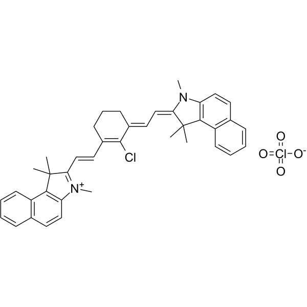
- Name: IR-813 perchlorate
- Chemical Name: ir-813 perchlorate
- CAS Number: 201024-57-9
- Molecular Formula: C36H38Cl2N2O4
- Molecular Weight: 633.60400
- Catalog: Research Areas Others
- Create Date: 2018-03-19 08:00:00
- Modify Date: 2024-01-03 01:44:34
-
IR 813 perchlorate is a near-infrared (NIR) fluorescent dye (Ex=815 nm, Em=840 nm) and can be used for visualizing regional lymph nodes in mice[1].
| Name | ir-813 perchlorate |
|---|---|
| Synonyms |
IR-813 perchlorate,2-[2-[2-Chloro-3-[2-(1,3-dihydro-1,1,3-trimethyl-2H-benzo[e]-indol-2-ylidene)-ethylidene]-1-cyclohexen-1-yl]-ethenyl]-1,1,3-trimethyl-1H-benzo[e]indolium perchlorate
2-[2-[2-chloro-3-[2-(1,3-dihydro-1,1,3-trimethyl-2h-benzo[e]-indol-2-ylidene)-ethylidene]-1-cyclohexen-1-yl]-ethenyl]-1,1,3-trimethyl-1h-benzo[e]indolium perchlorate |
| Description | IR 813 perchlorate is a near-infrared (NIR) fluorescent dye (Ex=815 nm, Em=840 nm) and can be used for visualizing regional lymph nodes in mice[1]. |
|---|---|
| Related Catalog | |
| In Vitro | IR 813 (17.3 μM, 2 h) induces a 31.4% hemolysis in red blood cells[1]. IR 813 (5.9 μM, 24 h) induces 50% cell death in MRC-5 cells[1]. |
| In Vivo | IR 813 perchlorate displays a maximum fluorescence intensity at 4 h postinjection together with a rapid extravasation, when being used for visualizing regional lymph nodes in mice[1]. Guidelines (Following is our recommended protocol. This protocol only provides a guideline, and should be modified according to your specific needs)[1]. 1. Animal: Ten-week-old female Balb/cOlaAnN mice are kept in a 12 h light/dark cycle to reduce tissue autofluorescence in the NIR region, and had access to food and water ad libitum. 2. Dose: A single dose 5.1 nmol of IR 813 dye (20 μL of 0.173 mg/mL, dissolved in a PEG-400/ethanol/water=3:2:5, v/v/v solution) is subcutaneous injected in the right anterior paw of mice. 3. Imaging: Using the Fluobeam700 NIR imaging system to perform in vivo optical imaging (739 nm excitation light (3.5 mW), 750 nm long-pass emission cutoff filter).Fluorescence intensity in the axillary lymph node (ALN) is recorded for 1 week (5 min, 1 h, 4 h, 24 h, 7 days). 4. Data analysis: Semiquantitative data is obtained from the fluorescence images using ImageJ 1.44 software. |
| References |
| Melting Point | 238-242ºC |
|---|---|
| Molecular Formula | C36H38Cl2N2O4 |
| Molecular Weight | 633.60400 |
| Exact Mass | 632.22100 |
| PSA | 80.52000 |
| LogP | 9.55480 |
| Personal Protective Equipment | Eyeshields;Gloves;type N95 (US);type P1 (EN143) respirator filter |
|---|---|
| Safety Phrases | S22-S24/25 |