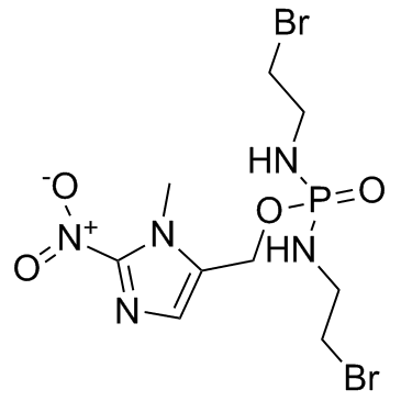
918633-87-1
| Name | 2-bromo-N-[(2-bromoethylamino)-[(3-methyl-2-nitroimidazol-4-yl)methoxy]phosphoryl]ethanamine |
|---|---|
| Synonyms |
(1-Methyl-2-nitro-1H-imidazol-5-yl)methyl N,N'-bis(2-bromoethyl)phosphorodiamidate
evofosfamide Phosphorodiamidic acid, N,N'-bis(2-bromoethyl)-, (1-methyl-2-nitro-1H-imidazol-5-yl)methyl ester cc-588 HAP-302 UNII-8A9RZ3HN8W TH-302 |
| Description | Evofosfamide (TH-302) is a hypoxia-activated prodrug with IC50 of 10 μM and 1000 μM in hypoxia (N2) and normoxia (21% O2), respectively. |
|---|---|
| Related Catalog | |
| Target |
Hypoxia-activated prodrug[1] |
| In Vitro | Evofosfamide (TH-302) induces γH2AX and apoptosis. Evofosfamide displays hypoxia-selective and concentration-dependent cytotoxic activity that is comparable in both p53-proficient and -deficient cells. Treatment with Evofosfamide (TH-302) alone causes an accumulation of G2/M cells. Inhibition of Chk1 by PF47736 in cells treated with Evofosfamide reduces Evofosfamide (TH-302)-mediated G2/M arrest under both normoxia and hypoxia[1]. |
| In Vivo | Evofosfamide (TH-302) is a hypoxia-activated prodrug known to activate selectively under the hypoxic conditions commonly found in solid tumors. The mean values of normalized Ktrans decrease 69.2% for Evofosfamide (TH-302)-treated mice in Hs766t tumors, decrease 46.1% for Mia PaCa-2 tumors and increase 4.9% in SU.86.86 tumors. Both changes for Hs766t and Mia PaCa-2 treatment groups are statistically significant (P<0.01) when compare to their own control group[2]. A significant reduction in the hypoxic fraction (HF) to 2.1%±4.7% is seen after 95% oxygen breathing (P<0.001), whereas 7% oxygen breathing significantly increase the HF to 29.5%±14.7% (P=0.029). Exposing rhabdomyosarcoma-bearing rats to increasing oxygen conditions abolish the effect of TH-302 and reduce the T4×SV from 20.4±3.5 to 15.3±2.5 days (P=0.007), whereas control animals have an increased T4×SV. Upon combination with radiotherapy, the T4×SV of TH-302-treated tumors decrease from 30.8±5.9 (Evofosfamide (TH-302)+radiotherapy) to 25.7±2.9 days (Evofosfamide (TH-302)+radiotherapy+95% O2)[3]. |
| Cell Assay | Cells are treated with 0.1 μM of either PF477736 or AZD7762 and Evofosfamide (TH-302) for 2 h under either normoxia (21% O2) or hypoxia (N2). Following wash, cells are cultured for additional 22 h in the presence of Chk1 inhibitor under normoxia. Cells are fixed in 75% ethanol and cell cycle distribution is determined using cell cycle reagent and Guava flow cytometry. HT-29 cells are exposed to Evofosfamide (TH-302)e (8 nM, 40 nM, 200 nM, 1 μM, and 5 μM) and 0.1 μM of AZD7762 for 2 h under either normoxia (21% O2) or hypoxia (N2). After wash, cells are continuously cultured for additional 46 h in the presence of 0.1 μM of AZD7762. Luminescence-based caspase activity assay is performed[1]. |
| Animal Admin | Mice[2] Female SCID mice of age 5-6 weeks are inoculated with SU.86.86, Hs766t or Mia-PaCa2 cells (5×10⁶) subcutaneously on the left hind leg. Tumors are allowed to grow for an average of three weeks to an average size of ~150 mm3. Mice are then randomized and placed into cohorts and treated with saline (control) or Evofosfamide (TH-302) (50 mg/kg) injected intraperitoneally. A total of 34 mice underwent MR imaging studies. The SU.86.86 group consist of 5 TH-302 treated and 5 control animals; Mia-PaCa2 consist of 6 Evofosfamide treated and 5 control animals; Hs766t consist of 7 Evofosfamide treated and 6 control animals. Animals are sacrificed when tumors reach 2000 mm3. Rats[2] Syngeneic rhabdomyosarcoma R1 tumors (1 mm3) are implanted subcutaneously in the lateral flank of adult WAG/Rij rats. Experiments are started upon a mean tumor volume of 4.2 cm3(range, 2.0-8.1) to ensure a stable HF. Treatment is administered on 4 consecutive days and consist of an intraperitoneal injection (i.p.; QD×4) with either NaCl or Evofosfamide (TH-302) (25, 50, or 75 mg/kg). Before the start of treatment, a PET scan is made using [18F]HX4. Radiotherapy is applied in a single dose of 0, 4, 8, or 12 Gy on day 3 of the treatment, 3 hours after NaCl or Evofosfamide (TH-302) injection, 1 hour after oxygen modification. During both PET imaging and radiotherapy, rats are anesthetized using a mixture of ketamine/xylazine (i.p; 66.7 and 6.7 mg/kg, respectively). During the 5 days of treatment (1 day PET imaging, 4 days of injections with Evofosfamide or vehicle), animals are exposed to modified oxygen concentrations for 4 hours per day in order to alter the HF of the tumor. The combination oxygen modification of nicotinamide (i.p. 500 mg/kg) and carbogen (95% oxygen, 5% CO2; 5 L/minute) consist of a nicotinamide injection and 30 minutes later the exposure to carbogen breathing for 3.5 hours. In the middle of the nicotinamide/carbogen treatment, NaCl/Evofosfamide is administered. Reduced oxygen breathing (7%, residual N2; 2.5 L/minute) is given for 4 hours with the NaCl/Evofosfamide injection after the first 2 hours. The injection of the [18F]HX4 PET tracer [mean 18.8 MBq, range 7.1-25.1 MBq; lateral tail vein using an intravenous line (Venoflux 0.4 mm G27) flushed with 10% heparine)] is given 2 hours before the end of the oxygen modification. PET imaging is performed 3 hours after tracer injection. |
| References |
| Density | 2.0±0.1 g/cm3 |
|---|---|
| Boiling Point | 565.4±60.0 °C at 760 mmHg |
| Molecular Formula | C9H16Br2N5O4P |
| Molecular Weight | 449.036 |
| Flash Point | 295.7±32.9 °C |
| Exact Mass | 446.930664 |
| PSA | 123.81000 |
| LogP | -0.40 |
| Vapour Pressure | 0.0±1.5 mmHg at 25°C |
| Index of Refraction | 1.662 |
| Storage condition | -20℃ |
