1207456-01-6
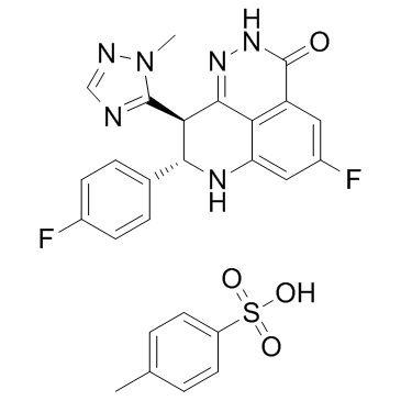
| Name | bmn 673 |
|---|---|
| Synonyms |
(8S,9R)-5-fluoro-8-(4-fluorophenyl)-9-(1-methyl-1H-1,2,4-triazol-5-yl)-8,9-dihydro-2H-pyrido[4,3,2-de]phthalazin-3(7H)-one
BMN-673 LT-673 3H-Pyrido[4,3,2-de]phthalazin-3-one, 5-fluoro-8-(4-fluorophenyl)-2,7,8,9-tetrahydro-9-(1-methyl-1H-1,2,4-triazol-5-yl)-, (8S,9R)- (8S,9R)-5-Fluoro-8-(4-fluorophenyl)-9-(1-methyl-1H-1,2,4-triazol-5-yl)-2,7,8,9-tetrahydro-3H-pyrido[4,3,2-de]phthalazin-3-one Talazoparib BMN673 |
| Description | Talazoparib (BMN-673) is a highly potent PARP1/2 inhibitor with Kis of 1.2 nM and 0.87 nM, respectively. |
|---|---|
| Related Catalog | |
| Target |
PARP2:0.87 nM (Ki) PARP1:1.2 nM (Ki) |
| In Vitro | Talazoparib (BMN 673) demonstrates excellent potency, inhibiting PARP1 and PARP2 enzyme activity with Ki=1.2 and 0.87 nM, respectively[1]. Talazoparib (BMN 673) exhibits selective antitumor cytotoxicity and elicits DNA repair biomarkers at much lower concentrations than earlier generation PARP1/2 inhibitors (such as Olaparib, Rucaparib, and Veliparib)[2]. |
| In Vivo | Talazoparib (BMN 673; 1 mg/kg, p.o.) is orally available, displaying favorable pharmacokinetic (PK) properties and remarkable antitumor efficacy in the BRCA1 mutant MX-1 breast cancer xenograft model following oral administration as a single-agent or in combination with chemotherapy agents such as temozolomide and cisplatin[1]. Talazoparib (BMN 673) is readily orally bioavailable, with more than 40% absolute oral bioavailability in rats when dosed in carboxylmethyl cellulose. Oral administration of Talazoparib elicits remarkable antitumor activity, xenografted tumors that carry defects in DNA repair due to BRCA mutations or PTEN deficiency are profoundly sensitive to oral Talazoparib treatment at well-tolerated doses in mice[2]. |
| Kinase Assay | The ability of a test compound to inhibit PARP1 enzyme activity is assessed using PARP1 assay kit. IC50 values are calculated using GraphPad Prism5 software. For PARP inhibitor Ki determination, enzyme assays are conducted in 96-well FlashPlate with 0.5 U of PARP1 enzyme, 0.25× activated DNA, 0.2 μCi [3H]NAD, and 5 μM cold NAD in a final volume of 50 μL in reaction buffer containing 10% glycerol (v/v), 25 mM HEPES, 12.5 mM MgCl2, 50 mM KCl, 1 mM dithiothreitol (DTT), and 0.01% NP-40 (v/v), pH 7.6. Reactions are initiated by adding NAD to the PARP reaction mixture with or without inhibitors and incubated for 1 min at room temperature. Fifty microliters of ice-cold 20% trichloroacetic acid (TCA) is then added to each well to quench the reaction. The plate is sealed and shaken for a further 120 min at room temperature, followed by centrifugation. Radioactive signal bound to the FlashPlate is determined using TopCount. PARP1 Km is determined using the Michaelis-Menten equation from various substrate concentrations (1-100 μM NAD). Compound Ki is calculated from the enzyme inhibition curve according to the following formula: Ki=IC50/[1+(substrate)/Km]. Km for PARP2 enzyme and compound Ki are determined with the same assay protocol except that 30 ng of PARP2, 0.25× activated DNA, 0.2 μCi [3H]NAD, and 20 μM cold NAD are used in the reaction for 30 min at room temperature[1]. |
| Cell Assay | In single-agent assays, Capan-1 cells (BRCA2-deficient), MX-1 (BRCA1-deficient) cells, or other cultured cells are seeded at densities that allow linear growth for 10-12 days in 96-well plates (typically 500-3000 cells/well). Cells are treated in their recommended growth media containing varying concentrations of PARP inhibitors (Veliparib, Rucaparib, Niraparib, Olaparib, and Talazoparib) for 10-12 consecutive days (media are changed with fresh compounds every 5 days). IC50 values are calculated using GraphPad Prism5[1]. |
| Animal Admin | Mice[1] MX-1 tumor xenografts are prepared. When tumors reach an average volume of approximately 150 mm3, Olaparib (100 mg/kg), Talazoparib (1 mg/kg), or vehicle is administered in a single po dose. Tumors are harvested at 2, 8, and 24 h after drug dosing and snap-frozen in liquid nitrogen. Tumor tissue is then homogenized in PBS on ice and extracted with lysis buffer (25 mM Tris, pH 8.0, 150 mM NaCl, 5 mM EDTA, 2 mM EGTA, 25 mM NaF, 2 mM Na3VO4, 1 mM Pefabloc, 1% Triton X-100, and protease inhibitor cocktail) containing 1% SDS. Levels of PAR in the tumor lysates are determined by ELISA using PARP in vivo PD assay II kit. |
| References |
| Density | 1.6±0.1 g/cm3 |
|---|---|
| Molecular Formula | C19H14F2N6O |
| Molecular Weight | 380.351 |
| Exact Mass | 380.119720 |
| PSA | 88.75000 |
| LogP | 1.91 |
| Index of Refraction | 1.775 |
| Hazard Codes | Xi |
|---|
| Precursor 8 | |
|---|---|
| DownStream 0 | |


![1(3H)-Isobenzofuranone,6-fluoro-3-[(1-Methyl-1H-1,2,4-triazol-5-yl)Methylene]-4-nitro-,(3Z)- structure](https://image.chemsrc.com/caspic/093/1322616-34-1.png)


