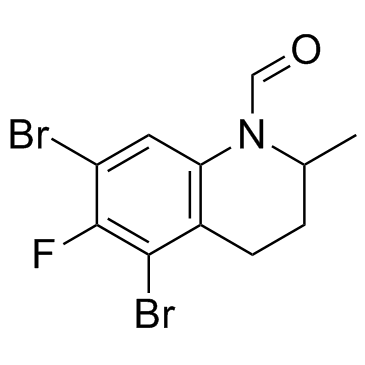
143703-25-7
| Name | CE3F4 |
|---|---|
| Synonyms |
1(2H)-Quinolinecarboxaldehyde, 5,7-dibromo-6-fluoro-3,4-dihydro-2-methyl-
5,7-Dibromo-6-fluoro-2-methyl-3,4-dihydro-1(2H)-quinolinecarbaldehyde CE3F4 |
| Description | CE3F4 is a selective antagonist of exchange protein directly activated by cAMP (Epac1), with IC50s of 10.7 μM and 66 μM for Epac1 and Epac2(B), respectively. |
|---|---|
| Related Catalog | |
| Target |
IC50: 10.7 μM (Epac1), 66 μM (Epac2(B))[1] |
| In Vitro | CE3F4 is a selective antagonist of Epac1, with IC50s of 10.7 μM and 66 μM for Epac1 and Epac2(B), respectively. CE3F4 is more active on Epac1 than (S)-stereoisomer ((S)-CE3F4, IC50, 56 μM), but less active than (R)-CE3F4 (IC50, 5.8 μM). CE3F4 (50 μM) shows more inhibitory activities against GEF activity of Epac1, than that of Epac2(AB) or Epac2(B)[1]. CE3F4 reduces the exchange activity of Epac1 induced by 007, with IC50 of 23 ± 3 μM. CE3F4 (40 μM) specifically inhibits Epac1 guanine nucleotide exchange activity without interference with Rap1 activity or Epac1-Rap1 interaction. CE3F4 has no influence on PKA activity. CE3F4 (20 μM) inhibits Epac-induced Rap1 activation in living cultured HEK293 cells[2]. CE3F4 (20 μM) significantly inhibits the late phase of ERK activation stimulated by glucose in INS-1 cells[3]. |
| Kinase Assay | To determine Epac1 exchange activity, 200 nM of purified GST-Rap1A preloaded with bGDP are incubated at 22°C in exchange buffer (50 mM Tris-HCl (pH 7.5), 50 mM NaCl, 5 mM MgCl2, 5 mM 1,4-dithioerythritol, 5% glycerol, 0.01% Nonidet P-40) in the presence of 100 nM purified GST-Epac1 or GST-Epac1-Cat; 20 μM unlabeled GDP; and defined concentrations of cAMP, cyclic nucleotide analogs, and test compounds (CE3F4). Experiments are performed in black 384-well plates in a final volume of 30 μL. bGDP fluorescence (excitation, 480 nm and emission, 535 nm) is measured using a multilabel plate reader[2]. |
| Cell Assay | Cells are cultured overnight in 96-well black-walled plates at 37°C and 5% CO2, then washed twice in phosphate buffered saline. Cells are pre-incubated for two hours in glucose-free, modified KRBH supplemented with 0.05% fatty acid-free BSA at 37°C and 5% CO2. The pre-incubation buffer is decanted, and cells are stimulated with 18 mM glucose in KRBH. Cells are incubated with or without inhibitors (CE3F4) in modified KRBH for 30 min at 37°C and 5 % CO2 before glucose stimulation. The reactions are terminated at the indicated time points by decanting the treatments and fixing the cells with 4% formaldehyde. In experiments using pharmacological inhibitors, reactions are terminated 10 min after glucose stimulation is initiated. Total ERK and pERK is measured using the Phospho-ERK1 (T202/Y204) / ERK2 (T185/Y187) Cell-Based ELISA. Total ERK1/ERK2 is measured at 450 nm with excitation at 360 nm, and phosphorylated ERK1/ERK2 is measured at 600 nm with excitation at 540 nm, using a Synergy 4 Microplate Reader. The data are expressed as the ratio of pERK to total ERK then normalized and expressed as either fold over basal or % glucose response[3]. |
| References |
| Density | 1.8±0.1 g/cm3 |
|---|---|
| Boiling Point | 431.1±45.0 °C at 760 mmHg |
| Molecular Formula | C11H10Br2FNO |
| Molecular Weight | 351.010 |
| Flash Point | 214.5±28.7 °C |
| Exact Mass | 348.911316 |
| LogP | 3.70 |
| Vapour Pressure | 0.0±1.0 mmHg at 25°C |
| Index of Refraction | 1.641 |
