| In Vitro |
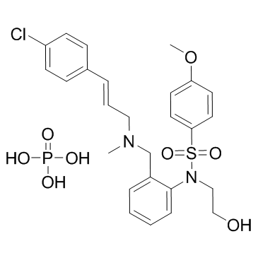
After 2 days of KN-93 treatment, 95% of cells are arrested in G1. G1 arrest is reversible; 1 day after KN-93 release, a peak of cells had progressed into S and G2-M. KN-93 also blocks cell growth stimulated by basic fibroblast growth factor, platelet-derived growth factor-BB, epidermal growth factor, and insulin-like growth factor-1 in NIH 3T3 fibroblasts[1]. KN-93 inhibits the H+, K+-ATPase activity but strongly dissipates the proton gradient formed in the gastric membrane vesicles and reduces the volume of luminal space[2]. KN-93 (0.5 μM) prevents increased LV developed pressure during action potential prolongation and early afterdepolarizations. Ca2+-independent CaM kinase activity is increased during early afterdepolarizations and this increase is prevented by KN-93[3].
|
| Kinase Assay |
Cells are grown on 12-mm diameter glass coverslips in DMEM 100% serum and various concentrations of KN-93 or KN-92. After 0, 1, 2, and 3 days of culture in the presence of drug, coverslips are removed from culture, rinsed once in PBS, and then submerged in 100% methanol at -20°C for 3 min. Fixed cells are stored in PBS until staining using the TUNEL assay. Cells are overlaid on 20 μL PBS/1 mg/mL BSA for 30 min, rinsed in PBS, and then overlaid on 20 μL containing 100 mM sodium cacodylate (pH 6.8), 1 mM CoCl2, 0.1 mM DTT, 0.1 mg/mL BSA, 20 μM fluorescein-12-dUTP, and 0.1 unit/μL terminal transferase at 37°C for 60 min. Coverslips are rinsed in PBS twice, mounted on slides, and photographed using an OLYMPUS BX5O epifluorescent microscope using a UPLAN APO 40X oil immersion objective.
|
