| In Vitro |
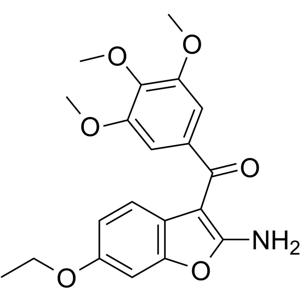
Tubulin polymerization-IN-13 (0.005-2.8 nM) treatment inhibits tumor cell proliferation[1]. Tubulin polymerization-IN-13 (8.7-10 μM) is non-toxic in non-tumor cells[1]. Tubulin polymerization-IN-13 (1-50 nM; 24 h) treatment induces cell cycle arrest in G2/M[1]. Tubulin polymerization-IN-13 (10 nM; 24 and 48 h) treatment induces cell apoptosis[1]. Tubulin polymerization-IN-13 (10-100 nM; 24 h) treatment induces alteration of the microtubule network[1]. Cell Proliferation Assay[1] Cell Line: HeLa, HT-29, Daoy, HL-60, SEM, and Jurkat cells Concentration: 0.005-2.8 nM Incubation Time: Result: Showed IC50s of 2.8 nM, 2.1 nM, 0.005 nM, 2.7 nM, 0.31 nM, and 0.28 nM for HeLa, HT-29, Daoy, HL-60, SEM, and Jurkat cells, respectively. Cell Cytotoxicity Assay[1] Cell Line: Peripheral blood lymphocytes (PBL) Concentration: 8.7-10 μM Incubation Time: Result: Showed a GI50 of 8.7 μM in quiescent lymphocytes. Cell Cycle Analysis[1] Cell Line: HeLa cells Concentration: 1, 5, 10, and 50 nM Incubation Time: 24 hours Result: Induced a G2/M arrest at 10 nM, increased G2/M cells accompanied by a strong reduction of cells in the G1 phase. Apoptosis Analysis[1] Cell Line: HeLa cells Concentration: 10 nM Incubation Time: 24 and 48 hours Result: Induced caspase-9 activation, PARP cleavage, Bcl-2 phosphorylation and Mcl-1 downregulation. Immunofluorescence[1] Cell Line: HeLa cells Concentration: 10, 50, and 100 nM Incubation Time: 24 hours Result: Showed the disorganization of microtubules at 10 nM, and much more evident at 50 and 100 nM.
|
