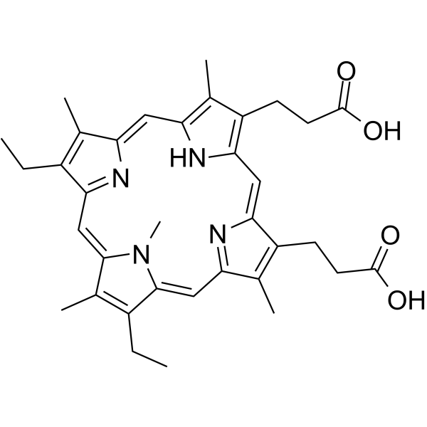
142234-85-3
| Name | 3-[18-(2-carboxyethyl)-7,12-diethyl-3,8,13,17,22-pentamethyl-23H-porphyrin-2-yl]propanoic acid |
|---|---|
| Synonyms |
N-methyl mesoporphyrin IX
21H,23H-porphine-2,18-dipropanoic acid,8,13-diethyl-3,7,12,17,23-pentamethyl |
| Description | N-Methylmesoporphyrin IX (NMM), a widely used G-quadruplex DNA specific fluorescent binder, is an efficient probe for monitoring Aβ fibrillation. N-Methylmesoporphyrin IX is an in situ inhibitor and an ex situ monitor for Aβ amyloidogenesis both in vitro and in cells. N-Methylmesoporphyrin IX is sensitive to G-quadruplexes DNA but has no response to duplexes, triplexes and single-stranded forms DNA. N-Methylmesoporphyrin IX is nonfluorescent alone or in monomeric Aβ environments, but emits strong fluorescence through stacking with the Aβ assemblies[1]. |
|---|---|
| Related Catalog | |
| In Vitro | By use of N-Methylmesoporphyrin IX (NMM) as an ex situ probe, NMM is added into the incubated Aβ40 solution. The concentration of NMM is fixed at 1μM. To examine the influence of Rhodamine B, 1 μM NMM with 0.1 μM or 0.5 μM Rhodamine B along with 10μM Aβ40 are measured. In the study of using NMM as an in situ inhibitor, NMM (10 μM) is co-incubated with Aβ40 (50 μM) for 7 days at 37 °C. Additional NMM is added before PL measurement to make the concentration of NMM constant (1 μM). The fluorescence spectra of NMM are collected from 550 to 700 nm with an excitation wavelength of 399 nm[1]. N-Methylmesoporphyrin IX (NMM) is sensitive to G-quadruplexes DNA but has no response to duplexes, triplexes and single-stranded forms DNA. Upon binding to quadruplex DNA, for efficient π-π stacking, NMM can adjust its macrocycle geometry to match the terminal face of a G-quadruplex, leading to an enhancement in its fluorescence[1]. |
| References |
| Density | 1.26g/cm3 |
|---|---|
| Molecular Formula | C35H40N4O4 |
| Molecular Weight | 580.71600 |
| Exact Mass | 580.30500 |
| PSA | 120.04000 |
| LogP | 4.08550 |
| Index of Refraction | 1.642 |
| Hazard Codes | Xi |
|---|
