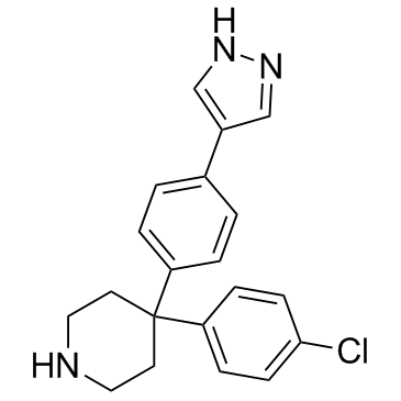
857531-00-1
| Name | 4-(4-Chlorophenyl)-4-[4-(1H-pyrazol-4-yl)phenyl]piperidine |
|---|---|
| Synonyms |
GVP
4-(4-(1H-pyrazol-4-yl)phenyl)-4-(4-chlorophenyl)piperidine AT7867 S1558_Selleck Piperidine, 4-(4-chlorophenyl)-4-[4-(1H-pyrazol-4-yl)phenyl]- AT-7867,AT7867 4-(4-chlorophenyl)-4-(4-(1h-pyrazol-4-yl)phenyl)piperidine 4-(4-Chlorophenyl)-4-[4-(1H-pyrazol-4-yl)phenyl]piperidine |
| Description | AT7867 is a potent ATP-competitive inhibitor of Akt1/Akt2/Akt3 and p70S6K/PKA with IC50s of 32 nM/17 nM/47 nM and 85 nM/20 nM, respectively. |
|---|---|
| Related Catalog | |
| Target |
Akt2:17 nM (IC50) Akt1:32 nM (IC50) Akt3:47 nM (IC50) PKA:20 nM (IC50) |
| In Vitro | The inhibition of AKT2 by AT7867 is shown to be ATP-competitive with a Ki of 18nM. AT7867 also displays potent activity against the structurally related AGC kinases p70S6K and PKA, but shows a clear window of selectivity against kinases from other kinase sub-families. In vitro growth inhibition studies show that AT7867 blocks proliferation in a number of human cancer cell lines. AT7867 appears to be most potent at inhibiting proliferation in MES-SA uterine, MDA-MB-468 and MCF-7 breast, and HCT116 and HT29 colon lines (IC50 values range from 0.9-3 μM), and least effective in the two prostate lines tested (IC50 values range from 10-12 μM) [1]. |
| In Vivo | In vivo: Following oral administration at 20 mg/kg, the elimination of AT7867 from plasma appears to be similar to that observed after i.v. administration. Plasma levels of AT7867 remain above 0.5 μM for at least 6 hours following an oral dose of 20 mg/kg. Assuming linear pharmacokinetics following i.v. administration, the bioavailability by the oral route is calculated to be 44%. In vivo pharmacodynamic (PD) biomarker studies are therefore performed with this model. Following pharmacokinetic and tolerability studies, doses of AT7867 (90 mg/kg p.o. or 20 mg/kg i.p.) are administered to athymic mice bearing MES-SA tumors and the phosphorylation status of GSK3β and S6RP in tumors is monitored over time. Clear inhibition of phosphorylation of the two markers of pathway activity is seen at 2 and 6 hours following treatment with AT7867. By 24 hours, total levels of both GSK3β and S6RP are greatly reduced[1]. |
| Kinase Assay | Kinase assays for AKT2, PKA, p70S6K and CDK2/cyclinA are all carried out in a radiometric filter binding format. Assay reactions are set up in the presence of compound. For AKT2, the AKT2 enzyme and 25 μM AKTide-2T peptide (HARKRERTYSFGHHA) are incubated in 20 mM MOPS, pH 7.2, 25 mM β-glycerophosphate, 5 mM EDTA, 15 mM MgCl2, 1 mM sodium orthovanadate, 1 mM DTT, 10 μg/mL BSA and 30 μM ATP (1.16 Ci/mmol) for 4 hours. For PKA, the PKA enzyme and 50 μM peptide (GRTGRRNSI) are incubated in 2 mM MOPS, pH 7.2, 25 mM β-glycerophosphate, 5 mM EDTA, 15 mM MgCl2, 1 mM orthovanadate, 1 mM DTT and 40 μM ATP (0.88 Ci/mmol) for 20 minutes. For p70S6K, the p70S6K enzyme and 25 μM peptide substrate (AKRRRLSSLRA) are incubated in 10 mM MOPS, pH 7, 0.2 mM EDTA, 1 mM MgCl2, 0.01% β-mercaptoethanol, 0.1 mg/mL BSA, 0.001% Brij-35, 0.5% glycerol and 15μM ATP (2.3 Ci/mmol) for 60 minutes. For CDK2, the CDK2/cyclinA enzyme and 0.12 μg/ml Histone H1 are incubated in 20 mM MOPS, pH 7.2, 25 mM β-glycerophosphate, 5 mM EDTA, 15 mM MgCl2, 1 mM sodium orthovanadate, 1 mM DTT, 0.1 mg/ml BSA and 45 μM ATP (0.78 Ci/mmol) for 4 hours. Assay reactions are stopped by adding an excess of orthophosphoric acid and the stopped reaction mixture is then transferred to Millipore MAPH filter plates and filtered. The plates are then washed, scintillant added and radioactivity measured by scintillation counting on a Packard TopCount. IC50 values are calculated from replicate curves using GraphPad Prism software. AKT1 and 3 enzyme assays are carried out, while all other enzyme assays are performed[1]. |
| Cell Assay | Cells are plated in 96-well microplates at 16,000 cells per well in medium supplemented with 10% FBS, and grown for 24 hours before treatment with AT7867. AT7867 or vehicle control are added to the cells for 1 hour. Following this, cells are fixed with 3% paraformaldehyde, 0.25% glutaraldehyde, 0.25% Triton-X100, washed and blocked with 5% milk in tris-buffered saline with 0.1% Tween-20 (TBST) prior to overnight incubation with a phospho-GSK3β (serine 9) antibody. The plates are then washed, secondary antibody added, and enhancement of the signal performed using DELFIA reagents. Europium counts are normalized to the protein concentration, and the IC50 value for each inhibitor is calculated in GraphPad Prism using non-linear regression analysis and a sigmoidal dose-response (variable slope) equation[1]. |
| Animal Admin | Mice[1] Male athymic BALB/c mice (nu/nu) are used. A single dose of AT7867 is administered to BALB/c mice at 5 mg/kg intravenously (i.v.) and 20 mg/kg per os (p.o.). Plasma samples are collected from duplicate animals at each of the following time points; 0.083, 0.167, 0.33, 0.67, 1, 2, 4, 6, 16 and 24 hours following i.v. dosing and at 0.25, 0.5, 1, 2, 4, 6 and 24 hours following p.o. dosing. Mice are bled by cardiac puncture and all blood samples are centrifuged to obtain plasma, which is then frozen at -20°C until analysis. For bioanalysis, all plasma samples are prepared by protein precipitation with acetonitrile containing internal standard. Quantification of sample extracts is by comparison with a standard calibration line constructed with AT7867 and using an inhibitor specific liquid chromatography tandem mass spectrometry (LC-MS/MS) method. Pharmacokinetic parameters are determined. |
| References |
| Density | 1.2±0.1 g/cm3 |
|---|---|
| Boiling Point | 530.2±50.0 °C at 760 mmHg |
| Molecular Formula | C20H20ClN3 |
| Molecular Weight | 337.846 |
| Flash Point | 274.5±30.1 °C |
| Exact Mass | 337.134583 |
| PSA | 40.71000 |
| LogP | 4.41 |
| Vapour Pressure | 0.0±1.4 mmHg at 25°C |
| Index of Refraction | 1.616 |
| Storage condition | -20°C |
