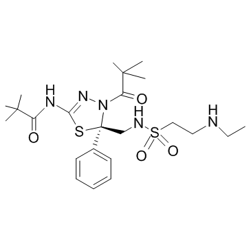
910634-41-2
| Name | N-[(5R)-4-(2,2-dimethylpropanoyl)-5-[[2-(ethylamino)ethylsulfonylamino]methyl]-5-phenyl-1,3,4-thiadiazol-2-yl]-2,2-dimethylpropanamide |
|---|---|
| Synonyms |
Litronesib (USAN/INN)
Litronesib [USAN] Litronesib UNII-6611F8KYCV |
| Description | Litronesib is a selective Eg5 inhibitor, with antitumor activity. |
|---|---|
| Related Catalog | |
| Target |
Eg5 |
| In Vitro | Litronesib (LY2523355) is a selective Eg5 inhibitor. Litronesib (25 nM) induces cancer cells to death during mitotic arrest, and this needs sustained activation of spindle-assembly checkpoint (SAC)[1]. |
| In Vivo | Litronesib (LY2523355; 1.1, 3.3, 10, and 30 mg/kg, i.v.) shows antitumor activity in a dose-dependently, and causes a dramatic increase in cancer cells immuno-positive for histone H3 phosphorylation in Colo205 xenograft tumors[1]. |
| Cell Assay | Cancer cells are plated in poly-d-lysine coated 96-well plates and incubated overnight. Cells are then treated with indicated concentrations of Litronesib for various time periods. Cells are then fixed with 3.7% formaldehyde in PBS for 45 minutes or 1× Prefer fixative solution for 30 minutes, at room temperature, and permeabilized with cold methanol for 10 minutes and then 0.2% Triton X-100 in PBS for 10 minutes. Cells are washed three times with PBS. Cells are then incubated with 100 μg/mL DNase-free RNase and 10 μg/mL of propidium iodide for 1 hour and scanned for mitotic index (MI) measurement based on DNA condensation as percentage of cells with condensed DNA. For MI based on histone H3 phosphorylation or apoptosis analysis, cells are incubated overnight at 4°C with anti-phospho-histone H3 antibody or anti-phospho-histone H2AX at 1:1,000 dilution with 5% bovine serum albumin (BSA) in PBS, respectively. Cells are washed three times with 0.2% Triton-X 100 in PBS and incubated with Alexa 488 secondary antibody (1:1,000 in PBS-2% BSA) for 60 minutes at room temperature. Cells are washed three times again with PBS and stained for 15 minutes in PBS containing 10 μg/mL propidium iodide and 100 μg/mL RNaseA. The stained cells are scanned with an Acumen Explorer eX3 microplate cytometer. The results are expressed as percentage of cells positive for phospho-histone H3-Ser10 or phospho-histone H2AX[1]. |
| Animal Admin | Primary human-tumor xenograft models are established and maintained in nude mice. Antitumor efficacy in subcutaneous xenograft tumor-bearing mice with 10 mice per treatment group from either established cancer cell lines or fragments of human tumor explants is evaluated as tumor volume by serial caliper measurements and is calculated. The p388 syngeneic tumor model is developed for high-content imaging analysis using female BDF1 mice, weighing 20 to 23 g, which are acclimated in-house for one week before their use in experiments. p388 murine lymphocytic leukemia cells are authenticated by STR assay and maintained in RPMI1640 medium containing 10% FBS. For inoculation, the cells are washed with serum-free medium three times and 1.25 million cells are implanted by intraperitoneal injection into mice. On day 5 after implantation, mice are treated with Litronesib, via either intravenous bolus or intravenous infusion at appropriate doses and durations. Mice are euthanized and the ascitic (intraperitoneal) fluid containing the p388 tumor cells is drawn and analyzed by acumen, flow cytometry, and TUNEL assays for phospho-histone H3, G2-M, and apoptosis. For pharmacokinetic study, the blood samples are collected via cardiac puncture and generated plasma with EDTA and determined Litronesib exposure in the plasma[1]. |
| References |
| Density | 1.23 g/cm3 |
|---|---|
| Molecular Formula | C23H37N5O4S2 |
| Molecular Weight | 511.70100 |
| Exact Mass | 511.22900 |
| PSA | 157.14000 |
| LogP | 4.49950 |
| Storage condition | 2-8℃ |
