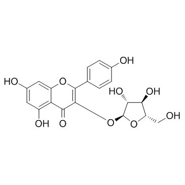
5041-67-8
| Name | Juglanin |
|---|---|
| Synonyms |
Juglanin
Kaempferol 3-O-arabinoside 5,7,4'-Trihydroxyflavone 3-α-L-arabinofuranoside 5,7-Dihydroxy-2-(4-hydroxyphenyl)-4-oxo-4H-chromen-3-yl α-L-arabinofuranoside 4H-1-Benzopyran-4-one, 3-(α-L-arabinofuranosyloxy)-5,7-dihydroxy-2-(4-hydroxyphenyl)- Euglanin Kaempferol 3-O-α-L-arabinofuranoside |
| Description | Juglanin is a JNK activator. |
|---|---|
| Related Catalog | |
| Target |
JNK |
| In Vitro | Juglanin inhibits the proliferation of breast cancer cell in a dose- and time-dependent manner and presents less cytotoxic against the normal cells. Juglanin results in the accumulation of cancer cells in G2/M phase and a corresponding decrease in G0/G1 and S phases in both MCF-7 and SKBR3 cells. Juglanin up-regulates the expressions of phosphorylated Chk2, Chk2, phosphorylated Cdc25C, phosphorylated Cdc2, p27, cyclin D and down-regulates the levels of Cdc25C as well as Cdc2. The proportion of apoptosis is negligible for control cells, whereas 24 h of exposure of cells to Juglanin leads to a dose-dependent increase of apoptotic cells in both MCF-7 and SKBR3 cells. Juglanin significantly suppresses anti-apoptotic factor of Bcl-2 expression, and in contrast Bad and Bax are both up-regulated for Juglanin treatment. Juglanin markedly activates caspases and leads to Casapse-9, Casapse-8 and Caspase-3 cleavage. ROS generation is initiated by Juglanin and significantly increased by high concentrations of Juglanin. Juglanin also increases the level of JNK phosphorylation in both MCF-7 and SKBR3 cells[1]. |
| In Vivo | Juglanin at doses of 5 and 10 mg/kg leads to significant decrease in tumor volume after 7 days of drug administration. 5 and 10 mg/kg Juglanin treatments do not cause weight loss in mice[1]. |
| Cell Assay | Breast cancer cells are planted in 96-well plates with a density of 5×10³ cells/well. After 12 h, the cells are treated with different concentrations of Juglanin (0 to 40 μM) for different periods of time (24 h and 48 h). Then the fresh mixture of MTS and PMS is added and incubated for 4 h at 37°C according to the manufacturer's protocol. A microplate reader is conducted to determine the absorbance at 500 nm, and the IC50 values are assessed with the probit model. Each one is performed in triplicate[1]. |
| Animal Admin | Male BALB/c-nude mice, aged 5 weeks, are used. They are maintained under specific pathogenfree conditions and supplied with sterilized food and water. On day 0, about 5×10⁶ MCF-7 cells suspended in 0.1 mL PBS are inoculated subcutaneously in the right flank of each mouse (six mice each group). On day 9, when the tumors reach palpable size of around 200 mm3, mice are randomly assigned to three groups and receive daily intraperitoneal injection with 100 μL of vehicle (10% DMSO, 70% Cremophor/ethanol (3: 1), and 20% PBS), and 5 or 10 Juglanin mg/kg of celastrol. Tumor sizes are measured daily to observe dynamic changes in tumor growth. Body weights are also measured daily. After 7 days of drug administration, when the tumors of control group reach around 1600 mm3, all mice are killed. Tumors are dissected and stored in liquid nitrogen or fixed in formalin for further analysis[1]. |
| References |
| Density | 1.8±0.1 g/cm3 |
|---|---|
| Boiling Point | 803.5±65.0 °C at 760 mmHg |
| Melting Point | 230-231℃ (ethanol ) |
| Molecular Formula | C20H18O10 |
| Molecular Weight | 418.351 |
| Flash Point | 289.0±27.8 °C |
| Exact Mass | 418.089996 |
| PSA | 170.05000 |
| LogP | 2.26 |
| Vapour Pressure | 0.0±3.0 mmHg at 25°C |
| Index of Refraction | 1.771 |
| Storage condition | 2-8℃ |
| Precursor 0 | |
|---|---|
| DownStream 1 | |

