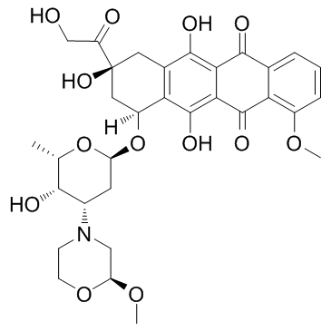
108852-90-0
| Name | nemorubicin |
|---|---|
| Synonyms |
(1S,3S)-3-Glycoloyl-3,5,12-trihydroxy-10-methoxy-6,11-dioxo-1,2,3,4,6,11-hexahydrotetracen-1-yl 2,3,6-trideoxy-3-[(2S)-2-methoxymorpholin-4-yl]-α-L-lyxo-hexopyranoside
Nemorubicin 5,12-Naphthacenedione, 7,8,9,10-tetrahydro-6,8,11-trihydroxy-8-(2-hydroxyacetyl)-1-methoxy-10-[[2,3,6-trideoxy-3-[(2S)-2-methoxy-4-morpholinyl]-α-L-lyxo-hexopyranosyl]oxy]-, (8S,10S)- (1S,3S)-3-Glycoloyl-3,5,12-trihydroxy-10-methoxy-6,11-dioxo-1,2,3,4,6,11-hexahydro-1-tetracenyl 2,3,6-trideoxy-3-[(2S)-2-methoxy-4-morpholinyl]-α-L-lyxo-hexopyranoside Nemorubicin [INN] (1S,3S)-3,5,12-trihydroxy-3-(hydroxyacetyl)-10-methoxy-6,11-dioxo-1,2,3,4,6,11-hexahydrotetracen-1-yl 2,3,6-trideoxy-3-[(2S)-2-methoxymorpholin-4-yl]-α-L-lyxo-hexopyranoside (1S,3S)-3-Glycoloyl-1,2,3,4,6,11-hexahydro-3,5,12-trihydroxy-10-methoxy-6,11-dioxo-1-naphthacenyl 2,3,6-Trideoxy-3-((S)-2-methoxymorpholino)-a-L-lyxo-hexopyranoside methoxy-morpholynil-doxorubicin |
| Description | Nemorubicin is a derivative of doxorubicin, and has antitumor activity. |
|---|---|
| Related Catalog | |
| In Vitro | Nemorubicin has antitumor activity, with IC70s of 578 ± 137 nM, 468 ± 45 nM, 193 ± 28 nM, 191 ± 19 nM, 68 ± 12 nM, and 131 ± 9 nM for HT-29, A2780, DU145, EM-2, Jurkat and CEM cell lines, respectively[1]. Nemorubicin acts through nucleotide excision repair (NER) system to exert its activity. Nemorubicin (0-0.3 μM) is more active in the L1210/DDP cells with intact NER than in the XPG-deficient L1210/0 cells. Cells resistant to nemorubicin show increased sensitivity to UV damage[3]. Nemorubicin is cytotoxic to 9L/3A4 cells, with an IC50 of 0.2 nM, 120-fold lower than that of P450-deficient 9L cells (IC50, 23.9 nM). Nemorubicin also potently inhibits Adeno-3A4 infected U251 cells with IC50 of 1.4 nM. P450 reductase overexpression enhances cytotoxicity of Nemorubicin[4]. |
| In Vivo | Nemorubicin is converted to PNU-159682 by human liver cytochrome P450 (CYP) 3A4 in rat, mouse, and dog liver microsomes[2]. Nemorubicin (60 µg/kg) induces sifnificant tumor growth delay in scid mice bearing 9L/3A4 tumors, but shows no obvious effect on the tumor growth delay of 9L tumors in mice by i.v. or intratumoral injection (i.t.). Nemorubicin (40 µg/kg, i.p.) exhibits no antitumor activity and no host toxicity in mice bearing 9L/3A4 tumors[4]. |
| Cell Assay | 9L and CHO cells are plated in triplicate wells of a 96-well plate at 3000 cells per well 24 hr prior to drug treatment. Cells are treated with various concentrations of Nemorubicin or IFA for 4d. Cells are then stained with crystal violet (A595) and relative cell survival is calculated. IC50 values are determined from a semi-logarithmic graph of the data points using Prism 4[4]. |
| Animal Admin | 9L and 9L/3A4 cells are grown as solid tumors in male ICR/Fox Chase SCID mice. Cells cultured in DMEM medium to 75% confluence are trypsinized and washed in PBS and then adjusted to 2 × 10⁷ cells/mL of FBS-free DMEM. Four-week-old SCID mice (18-20 g) are implanted with either 9L or 9L/3A4 tumor cells by injection of 4 × 10⁶ cells/0.2 mL of cell suspension, s.c. on each hind flank. Tumor sizes (length and width) are measured twice a week using Vernier calipers beginning 7d after tumor implantation. When the average tumor size reach 300 to 400 mm3, Nemorubicin dissolved in PBS is administered by tail vein injection (i.v.) or by direct intratumoral (i.t.) injection (three injections spaced 7 d apart, each at 60 µg Nemorubicin per kg body weight). Intratumoral injections are performed using a syringe pump set a 1 µL/s with a 30-gauge needle. Each i.t. treatment dose is divided into three injections per tumor, with the injected volume set at 50 µL per tumor per 25 g mouse. Thus, for a 30 g mouse, a total of 120 µL of 15 µg/mL of Nemorubicin solution is administered: 20 µL per site × 3 sites per tumor × 2 tumors/mouse. Drug-free controls are injected i.t. with the same vol of PBS. In some experiments, Nemorubicin is administered by i.p. injection at 40 or 60 µg/kg body weight. Tumor sizes and body weights are measured twice/wk for the duration of the study. Tumor volumes are calculated using the formula: V = π/6 (L × W)3/2. Percent tumor regression is calculated as 100 × (V1-V2)/V1, where V1 is the tumor vol on the day of drug treatment and V2 is the vol on the day when the largest the decrease in tumor size is seen following drug treatment. Tumor doubling time is calculated as the time required for tumors to double in vol after drug treatment[4]. |
| References |
| Density | 1.6±0.1 g/cm3 |
|---|---|
| Boiling Point | 852.2±65.0 °C at 760 mmHg |
| Molecular Formula | C32H37NO13 |
| Molecular Weight | 643.635 |
| Flash Point | 469.2±34.3 °C |
| Exact Mass | 643.226501 |
| PSA | 201.75000 |
| LogP | 4.70 |
| Vapour Pressure | 0.0±0.3 mmHg at 25°C |
| Index of Refraction | 1.681 |
| Storage condition | 2-8℃ |
| HS Code | 2933990090 |
|---|
| HS Code | 2933990090 |
|---|---|
| Summary | 2933990090. heterocyclic compounds with nitrogen hetero-atom(s) only. VAT:17.0%. Tax rebate rate:13.0%. . MFN tariff:6.5%. General tariff:20.0% |
