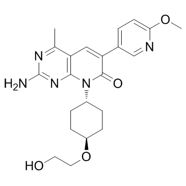
1013101-36-4
| Name | 2-amino-8-[4-(2-hydroxyethoxy)cyclohexyl]-6-(6-methoxypyridin-3-yl)-4-methylpyrido[2,3-d]pyrimidin-7-one |
|---|---|
| Synonyms |
2-Amino-8-[trans-4-(2-hydroxyethoxy)cyclohexyl]-6-(6-methoxy-3-pyridinyl)-4-methylpyrido[2,3-d]pyrimidin-7(8H)-one
2-amino-8-[trans-4-(2-hydroxyethoxy)cyclohexyl]-6-(6-methoxypyridin-3-yl)-4-methylpyrido[2,3-d]pyrimidin-7(8H)-one PF-04691502 Pyrido[2,3-d]pyrimidin-7(8H)-one, 2-amino-8-[trans-4-(2-hydroxyethoxy)cyclohexyl]-6-(6-methoxy-3-pyridinyl)-4-methyl- |
| Description | PF-04691502 is a potent and selective inhibitor of PI3K and mTOR. PF-04691502 binds to human PI3Kα, β, δ, γ and mTOR with Kis of 1.8, 2.1, 1.6, 1.9 and 16 nM, respectively. |
|---|---|
| Related Catalog | |
| Target |
PI3Kα:1.8 nM (Ki) PI3Kβ:2.1 nM (Ki) PI3Kδ:1.6 nM (Ki) PI3Kγ:1.9 nM (Ki) mTOR:16 nM (Ki) |
| In Vitro | PF-04691502 inhibits recombinant mouse PI3Kα in an ATP-competitive inhibitor. PF-04691502 potently inhibits AKT phosphorylation on S473 and T308 in all the 3 cancer cell lines with IC50 values of 3.8 to 20 nM and 7.5 to 47 nM, respectively. Using a 96-well plate-based P-S6RP(S235/236) ELISA assay, PF-04691502 potently inhibits mTORC1 activity with an IC50 of 32 nM. PF-04691502 inhibits cell proliferation of BT20, SKOV3, and U87MG with IC50 values of 313, 188, and 179 nM, respectively. In PIK3CA-mutant and PTEN-deleted cancer cell lines, PF-04691502 reduces phosphorylation of AKT T308 and AKT S473 (IC50 of 7.5-47 nM and 3.8-20 nM, respectively) and inhibits cell proliferation (IC50 of 179-313 nM). PF-04691502 inhibits mTORC1 activity in cells as measured by PI3K-independent nutrient stimulated assay, with an IC50 of 32 nM and inhibits the activation of PI3K and mTOR downstream effectors including AKT, FKHRL1, PRAS40, p70S6K, 4EBP1, and S6RP[1]. |
| In Vivo | Nude mice bearing U87MG tumors are administered orally once a day with PF-04691502 at 0.5, 1, 5, and 10 mg/kg (maximum tolerated dose, MTD). Treatment with 10 mg/kg results in a significant reduction of P-AKT(S473) levels at 1 hour postdosing, and persistent inhibition is observed for 8 hours. P-AKT(S473) recovers to above baseline 24 hours after 10 mg/kg treatment. For P-S6RP(S235/236), a similar inhibition time course is observed, but after 24 hours of treatment, P-S6RP levels remain lower than vehicle tumors. Modulation of the AKT downstream effector, P-PRAS40(T246), and mTOR downstream effector, P-4EBP1(T37/46), is observed. The PF-04691502-treated tumors are also evaluated by immunohistochemistry for levels of P-AKT(S473), total AKT, P-S6RP, and total S6RP. Phosphorylation of AKT and S6RP are significantly reduced at 4 hours after a single dose of PF-04691502 at 10 mg/kg. Dose-dependent tumor growth inhibition (TGI) is obtained in the U87MG xenograft model and approximately 73% TGI is observed at the MTD dose of 10 mg/kg[1]. |
| Kinase Assay | The biochemical protein kinase assays for class I PI3K and mTOR are assessed. The fluorescence polarization assay for ATP competitive inhibition is done as follows: mPI3Kα dilution solution (90 nM) is prepared in fresh assay buffer (50 mM Hepes pH 7.4, 150 mM NaCl, 5 mM DTT, 0.05% CHAPS) and kept on ice. The enzyme reaction contained 0.5 nM mouse PI3Kα (p110α/p85α complex purified from insect cells), 30 μM PIP2, PF-04691502 (0, 1, 4, and 8 nM), 5 mM MgCl2, and 2-fold serial dilutions of ATP (0-800 μM). Final DMSO is 2.5%. The reaction is initiated by the addition of ATP and terminated after 30 minutes with 10 mM EDTA. In a detection plate, 15 uL of detector/probe mixture containing 480 nM GST-Grp1PH domain and 12 nM TAMRA tagged fluorescent PIP3 in assay buffer is mixed with 15 uL of kinase reaction mixture. The plate is shaken for 3 minutes, and incubated for 35 to 40 minutes before reading on an LJL Analyst HT[1]. |
| Cell Assay | BT20, U87MG, and SKOV3 cells are plated at 3,000 cell/well in 96-well culture plates in growth medium with 10% FBS. Cells are incubated overnight and treated with DMSO (0.1% final) or serial diluted compound for 3 days. Resazurin is added to 0.1 mg/mL. Plates are incubated at 37°C in 5% CO2 for 3 hours. Fluorescence signals are read as emission at 590 nm after excitation at 530 nm. IC50 values are calculated by plotting fluorescence intensity to drug concentration in nonlinear curves. U87MG and SKOV3 cells are plated in 96-well plates overnight and caspase-3/caspase-7 activity is assessed with the Caspase-Glo 3/7 Assay Kit[1]. |
| Animal Admin | Mice[1] Female nu/nu mice (6-8 weeks old) are used. Tumor cells for implantation are harvested and resuspended in serum-free medium mixed with matrigel (1:1). SKOV3, U87MG, or NSCLC cells (2.5-4×10⁶) are implanted subcutaneously into the hind flank region. Treatment started when average tumor size is 100 to 200 mm3. PF-04691502 is formulated in 0.5% methylcellulose in water suspension and given orally once a day. Animal body weights and tumor volumes are measured every 2 to 3 days. Tumor volume is determined with Vernier calipers and calculated. Percentage of tumor growth inhibition (TGI) is calculated. Data are presented as mean±SE. Comparisons between treatment groups and vehicle group are done using 1-way ANOVA by Dunnett's tests. Student's t test is used to determine the P value for the comparison of 2 groups. |
| References |
| Density | 1.4±0.1 g/cm3 |
|---|---|
| Boiling Point | 682.5±65.0 °C at 760 mmHg |
| Molecular Formula | C22H27N5O4 |
| Molecular Weight | 425.481 |
| Flash Point | 366.5±34.3 °C |
| Exact Mass | 425.206299 |
| PSA | 125.38000 |
| LogP | 1.43 |
| Vapour Pressure | 0.0±2.2 mmHg at 25°C |
| Index of Refraction | 1.646 |
| Storage condition | -20℃ |
| RIDADR | NONH for all modes of transport |
|---|
