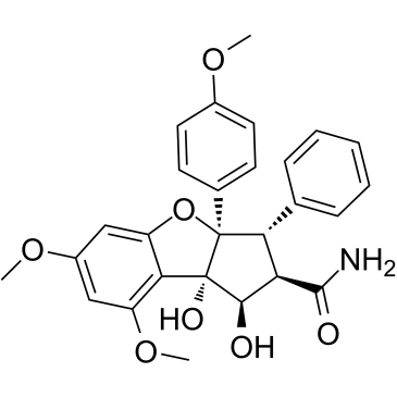
Didesmethylrocaglamide
Modify Date: 2024-01-10 22:54:29

Didesmethylrocaglamide structure
|
Common Name | Didesmethylrocaglamide | ||
|---|---|---|---|---|
| CAS Number | 177262-30-5 | Molecular Weight | 477.51 | |
| Density | N/A | Boiling Point | N/A | |
| Molecular Formula | C27H27NO7 | Melting Point | N/A | |
| MSDS | N/A | Flash Point | N/A | |
Use of DidesmethylrocaglamideDidesmethylrocaglamide, a derivative of Rocaglamide, is a potent eukaryotic initiation factor 4A (eIF4A) inhibitor. Didesmethylrocaglamide has potent growth-inhibitory activity with an IC50 of 5 nM. Didesmethylrocaglamide suppresses multiple growth-promoting signaling pathways and induces apoptosis in tumor cells. Antitumor activity[1]. |
| Name | Didesmethylrocaglamide |
|---|
| Description | Didesmethylrocaglamide, a derivative of Rocaglamide, is a potent eukaryotic initiation factor 4A (eIF4A) inhibitor. Didesmethylrocaglamide has potent growth-inhibitory activity with an IC50 of 5 nM. Didesmethylrocaglamide suppresses multiple growth-promoting signaling pathways and induces apoptosis in tumor cells. Antitumor activity[1]. |
|---|---|
| Related Catalog | |
| Target |
Eukaryotic initiation factor 4A (eIF4A)[1] |
| In Vitro | Didesmethylrocaglamide (5 nM, and 10 nM; 72 hours; MPNST cells) treatment arrests MPNST cells at G2-M, increases the sub-G1 population, induces cleavage of caspases and PARP, and elevates the levels of the DNA-damage response marker γH2A.X, while decreasing the expression of AKT and ERK1/2[1]. Didesmethylrocaglamide inhibits MPNST cell proliferation by inducing cell cycle arrest at G2/M and subsequently, cell death. Didesmethylrocaglamide-treated 697-R cells exhibits IC50 values is very similar to those of parental 697 cells (4 vs 3nM of IC50, respectively)[1]. Didesmethylrocaglamide induces apoptosis in both neurofibromatosis type 1 (NF1)-expressing and NF1-deficient MPNST cells, possibly subsequent to the activation of the DNA damage response. Didesmethylrocaglamide-treated sarcoma cells show decreased levels of multiple oncogenic kinases, including insulin-like growth factor-1 receptor[1]. Western Blot Analysis[1] Cell Line: Malignant peripheral nerve sheath tumors (MPNST) cells Concentration: 5 nM, and 10 nM Incubation Time: 72 hours Result: Induced cleavage of caspases and PARP, and elevated the levels of the DNA-damage response marker γH2A.X. |
| References |
| Molecular Formula | C27H27NO7 |
|---|---|
| Molecular Weight | 477.51 |
| Hazard Codes | Xi |
|---|