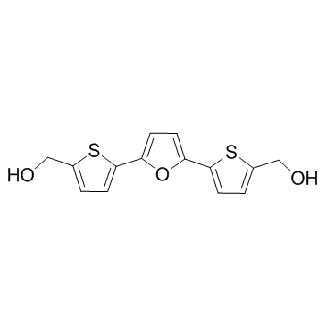
213261-59-7
| Name | [5-[5-[5-(hydroxymethyl)thiophen-2-yl]furan-2-yl]thiophen-2-yl]methanol |
|---|---|
| Synonyms |
p53 Activator III,RITA
(Furan-2,5-diyldithiene-5,2-diyl)dimethanol RITA (2,5-Furandiyldi-5,2-thienediyl)dimethanol 2-Thiophenemethanol, 5,5'-(2,5-furandiyl)bis- SOS BISMETHANOL Reactivation of p53 and Induction of Tumor cell Apoptosis NSC 652287 5,5'-(2,5-Furandiyl)bis-2-thiophenemethanol NSC652287 |
| Description | RITA is an inhibitor of p53-HDM-2 interaction, binds to p53dN, with a Kd of 1.5 nM, and also induces DNA-DNA cross-links. |
|---|---|
| Related Catalog | |
| Target |
Kd: 1.5 nM (p53dN)[1] DNA Crosslinker[2] |
| In Vitro | RITA inhibits p53-HDM-2 interaction, binding to p53dN, with a Kd of 1.5 nM. RITA (10 μM) blocks complex formation between p53 and HDM-2 in HCT116 cells and HDFs and in NHF-ERMyc cells irrespective of c-Myc expression. RITA (0.5 μM) reduces the viability of tumor cells in a wild-type p53-dependent manner. Moreover, RITA (0.1 μM) induces p53-dependent apoptosis. RITA induces p53 but does not via DNA damage-signaling pathway[1]. RITA (NSC 652287) induces DNA-DNA cross-links. RITA induces G2-M cell cycle arrest at 10 nM and causes apoptosis at 100 nM. RITA (100 nM) also elevates p53 and causes dose-dependent effects on p21WAF1 protein levels[2]. RITA inhibits the growth of HeLa and CaSki cells, with IC50s of 1 and 10 μM. In addition, RITA (1 μM) stabilizes p53 by inhibiting p53/E6AP interaction[3]. |
| In Vivo | RITA (0.1, 1 or 10 mg/kg, i.p.) shows potent antitumor activity in SCID mice bearing HCT116 and HCT116 TP53−/− xenografts[1]. RITA (10 mg/kg, i.p.) also suppresses the growth of HeLa cells in SCID mice[3]. |
| Cell Assay | For the cell viability assay, 3,000 cells per well are plated in a 96-well plate and treated with RITA for 48 h, after which cell viability is assessed with the proliferation reagent WST-1. For colony formation assay, cells are seeded in 12-well plates and treated with RITA for 24 h, after which the medium is replaced and the cells are allowed to grow for 10-14 d. The colonies are stained with crystal violet. For growth curves, 3000 cells/mL are plated in 12-well plates, treated with RITA, and counted over 5 d[3]. |
| Animal Admin | Mice[1] Female SCID mice, 4-6 weeks old, are implanted with subcutaneous xenografts using 1 × 10⁶ cells in 90% Matrigel. Palpable tumors are established 3-6 d after the cells are injected, at which point RITA treatment is initiated. RITA is administered either 0.1, 1 or 10 mg/kg every day by intravenous or intraperitoneal injection in a total volume of 100 μL phosphate buffered saline. Xenografts are measured every 2 d. Tumor volumes are plotted for control and treated groups by dividing the average tumor volume for each data point by average starting tumor volume[1]. |
| References |
| Density | 1.4±0.1 g/cm3 |
|---|---|
| Boiling Point | 464.9±40.0 °C at 760 mmHg |
| Melting Point | 160 °C |
| Molecular Formula | C14H12O3S2 |
| Molecular Weight | 292.373 |
| Flash Point | 235.0±27.3 °C |
| Exact Mass | 292.022797 |
| PSA | 110.08000 |
| LogP | 2.48 |
| Vapour Pressure | 0.0±1.2 mmHg at 25°C |
| Index of Refraction | 1.661 |
| Storage condition | Store at -20°C |
| Hazard Codes | Xi |
|---|
