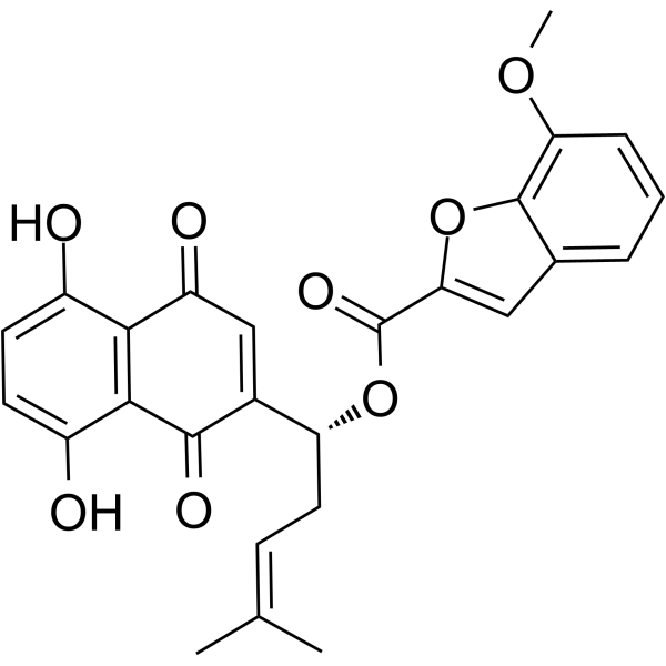
| In Vitro |
Tubulin inhibitor 25 (compound 6c) (0-100 μM; 48 hours) exhibits high antiproliferative activity against tested cancer cell lines[1]. Tubulin inhibitor 25 (0-200 μM; 48 hours) exhibits low cytotoxicity in normal cell lines[1]. Tubulin inhibitor 25 (0.05, 0.1 and 0.2 μM; 24 hours) inhibits the colony formation of HT29 cells in a dose-dependent manner[1]. Tubulin inhibitor 25 (2 and 4 μM) can inhibit the tubulin polymerization and compete with colchicine binding site[1]. Tubulin inhibitor 25 (0.25-1 μM; 12-48 hours) arrests the cell cycle at G2/M phase and induces HT29 cells apoptosis in a dose- and time-dependent manner, besides induces HT29 cell depolarized mitochondria in the process of apoptosis[1]. Tubulin inhibitor 25 (0.25-1 μM; 24 hours) increases the expression of P21 and Cyclin B1 and decreases the expression of Cdc2, p-CDC2 and p-Cdc25c; as well as induces the microtubule collapse in HT29 cells in a dose-dependent manner[1]. Tubulin inhibitor 25 (0.01, 0.02 and 0.04 μM; 6 hours) effectively inhibits the HUVEC tube formation in a dose-dependent manner[1]. Tubulin inhibitor 25 (0.1, 0.2 and 0.4 μM; 24 hours) inhibits migration of A549 cells in a dose-dependent manner[1]. Cell Proliferation Assay Cell Line: MDA-MB-231, HepG2, HT29, HCT116 and A549[1] Concentration: 0-100 μM Incubation Time: 48 hours Result: Exhibited high antiproliferative activity against HT29, HCT116, MDA-MB-231 and A549 with IC50s of 0.18 ± 0.04 μM, 0.58 ± 0.11 μM, 0.81 ± 0.13 μM and 0.57 ± 0.79 μM, and less activity against HepG2 with an IC50 of 73.20 ± 4.03 μM. Cell Cytotoxicity Assay Cell Line: 293T and LO2[1] Concentration: 0-200 μM Incubation Time: 48 hours Result: Exhibited low cytotoxicity in normal cell lines with CC50s of 184.86 ± 9.88 μM and 154.76 ± 9.98 μM in 293T and LO2. Cell Cycle Analysis Cell Line: HT29[1] Concentration: 0.25, 0.5 and 1 μM Incubation Time: 12, 24, 36 and 48 hours Result: Arrested the cell cycle at G2/M phase in a dose-dependent manner with the G2/M cell proportion of 23.05%, 23.55% and 80.99% at 0.25 μM, 0.5 μM and 1 μM, respectively, also exhibited time-dependent manner with the G2/M cell proportion of 32.55%, 36.43% and 71.1% for 12, 36 and 48 hours. Western Blot Analysis Cell Line: HT29[1] Concentration: 0.25, 0.5 and 1 μM Incubation Time: 24 hours Result: Increased the expression of P21 and Cyclin B1 and decreased the expression of Cdc2, p-CDC2 and p-Cdc25c.
|
