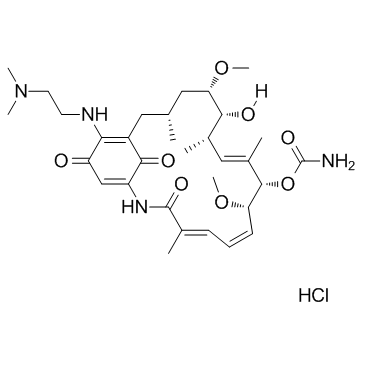
467214-21-7
| Name | [(3R,5S,6R,7S,8E,10S,11S,12Z,14E)-21-[2-(dimethylamino)ethylamino]-6-hydroxy-5,11-dimethoxy-3,7,9,15-tetramethyl-16,20,22-trioxo-17-azabicyclo[16.3.1]docosa-1(21),8,12,14,18-pentaen-10-yl] carbamate,hydrochloride |
|---|---|
| Synonyms |
17-N,N-Dimethylaminoethylamino-17-demethoxy-geldanamycin,HCl
17DMAG 17-DMAG (Alvespimycin) HCl Alvespimycin hydrochloride Alvespimycin hydrochloride (USAN) Alvespimycin HCl 17-Desmethoxy-17-N,N-dimethylaminoethylamino-geldanamycin,HCl Alvespimycin (hydrochloride) |
| Description | Alvespimycin hydrochloride is a potent inhibitor of Hsp90, binding to Hsp90 with EC50 of 62±29 nM. |
|---|---|
| Related Catalog | |
| Target |
HSP90:62 nM (EC50) GRP94:65 nM (EC50) |
| In Vitro | Alvespimycin (17-DMAG) hydrochloride inhibits the growth of the human cancer cell lines SKBR3 and SKOV3, which overexpress Hsp90 client protein Her2, and causes down-regulation of Her2 as well as induction of Hsp70 consistent with Hsp90 inhibition, for Her2 degradation with EC50 of 8±4 nM and 46±24 nM in SKBR3 and SKOV3 cells, respectively; for Hsp70 induction with EC50 of 4±2 nM and 14±7 nM in SKBR3 and SKOV3 cells, respectively[1]. Compared with the vehicle control, 17-DMAG exerts dose-dependent apoptosis (P<0.001 averaged across 24- and 48-hour time points) at concentrations of 50nM to 500nM, which represent pharmacologically attainable doses. Similar to many other agents, 17-DMAG also demonstrates time-dependent apoptosis (P <0.001, averaged across all doses) in chronic lymphocytic leukemia (CLL) cells with extended exposure from 24 to 48 hours. In addition, 17-DMAG is much more potent after 24 and 48 hours of treatment than 17-AAG[2]. |
| In Vivo | The tumors are grown for two months before the start of i.p. injections every four days over one month with 0, 50, 100 and 200 mg/kg dipalmitoyl-radicicol or 0, 5, 10 and 20 mg/kg 17-DMAG. Despite sample heterogeneity, the HSP90 inhibitor-treated animals have significantly lower tumour volumes than the vehicle control-treated animals. HSP90 inhibitors have been shown to cause liver toxicity in an animal model of gastrointestinal cancer. Nevertheless, the reduction in tumor size using dipalmitoyl-radicicol is statistically significant at 100 mg/kg, while 17-DMAG at either 10 or 20 mg/kg elicited a significant reduction in tumor size[3]. |
| Cell Assay | MTT assays are performed to determine cytotoxicity. A total of 1×10⁶ CD19-selected B cells from CLL patients are incubated for 24 or 48 hours in 17-DMAG, 17-AAG, or vehicle. MTT reagent is then added, and plates are incubated for an additional 24 hours before spectrophotometric measurement. Apoptosis is determined by staining with annexin V-fluorescein isothiocyanate and propidium iodide (PI). After exposure to drugs, cells are washed with phosphate-buffered saline and stained in 1 time binding buffer. Cell death is assessed by flow cytometry using a Beckman-Coulter model EPICS XL cytometer. Data are analyzed with the System II software package. A total of 10 000 cells are counted for each sample. Mitochondrial membrane potential changes are assessed by staining with the lipophilic cationic dye JC-1 and analysis by flow cytometry[2]. |
| Animal Admin | Mice[3] Young male CB-17/IcrHsd-Prkdc-SCID mice, are used. Recombinant xenografts are made by mixing 1×10⁵ BPH1 cells and 2.5×10⁵ CAF per graft in collagen solution, allowed to gel, covered with medium and cultured overnight. Tumors are allowed to form over eight weeks, and then treated for four weeks with three different doses of dipalmitoyl-radicicol (50, 100 and 200 mg/kg) and 17-DMAG (5, 10 and 20 mg/kg) via intraperitoneal injections of compounds in sesame oil every four days. After 12 weeks in total, the mice are sacrificed, their kidneys resected, grafts cut in half and photographed before processing for histology. Graft dimensions are measured and the resultant tumour volume is calculated using the formula; volume=width×length×depth×π/6. This formula represents a conservative approach to evaluate tumour volumes, as it understates the volume of large, invasive tumours compared with smaller, non-invasive tumours. Resected grafts are fixed in 10% formalin, embedded in paraffin and processed for immunohistochemistry. |
| References |
| Molecular Formula | C32H49ClN4O8 |
|---|---|
| Molecular Weight | 653.20600 |
| Exact Mass | 652.32400 |
| PSA | 174.00000 |
| LogP | 3.89580 |
| Storage condition | Store at -20 |
